Cutting:
Thinning:
Polishing:
Coating:
Cleaning:
Drying:
- Freeze dryer
- Critical point dryer (CPD)
The ultramicrotomy is a toll for sample preparation for different microscopic analysis methods, like TEM, SEM, AFM and LM. It generates ultrathin slices of material. Mostly this method is used on soft materials or biological samples. The section thickness varies between 10 nm and 500 nm.
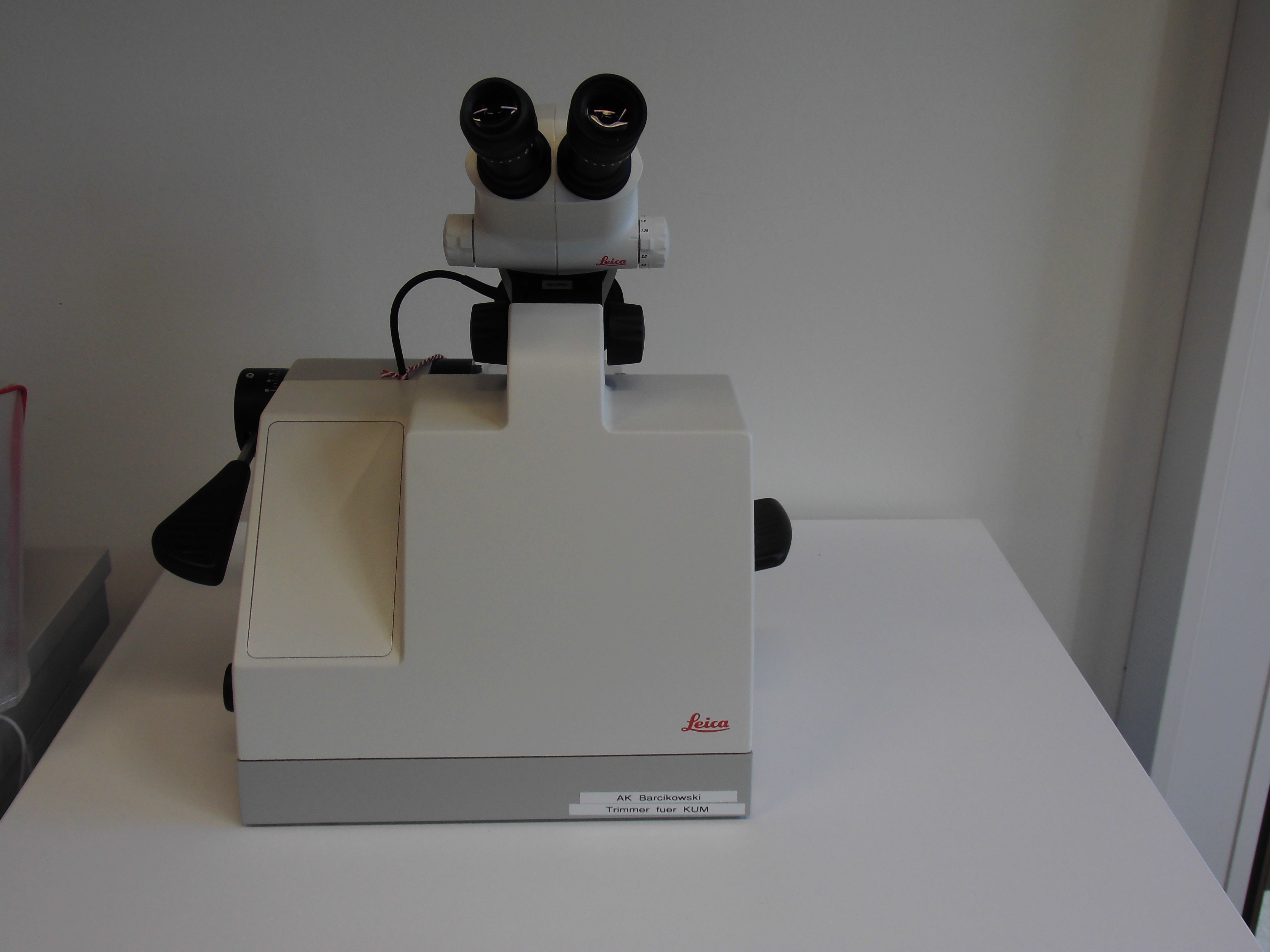
The sample is fixed in e.g. paraffin or epoxy before being cut. Usually these blocks have to trimmed prior to putting them in the microtome to achieve a perfect microtome sections. At the ICAN we use a EM TRIM2 by LEICA for trimming.
The 'blocks' are trimmed untiul the sample is clearly visible and a smooth surface is present.
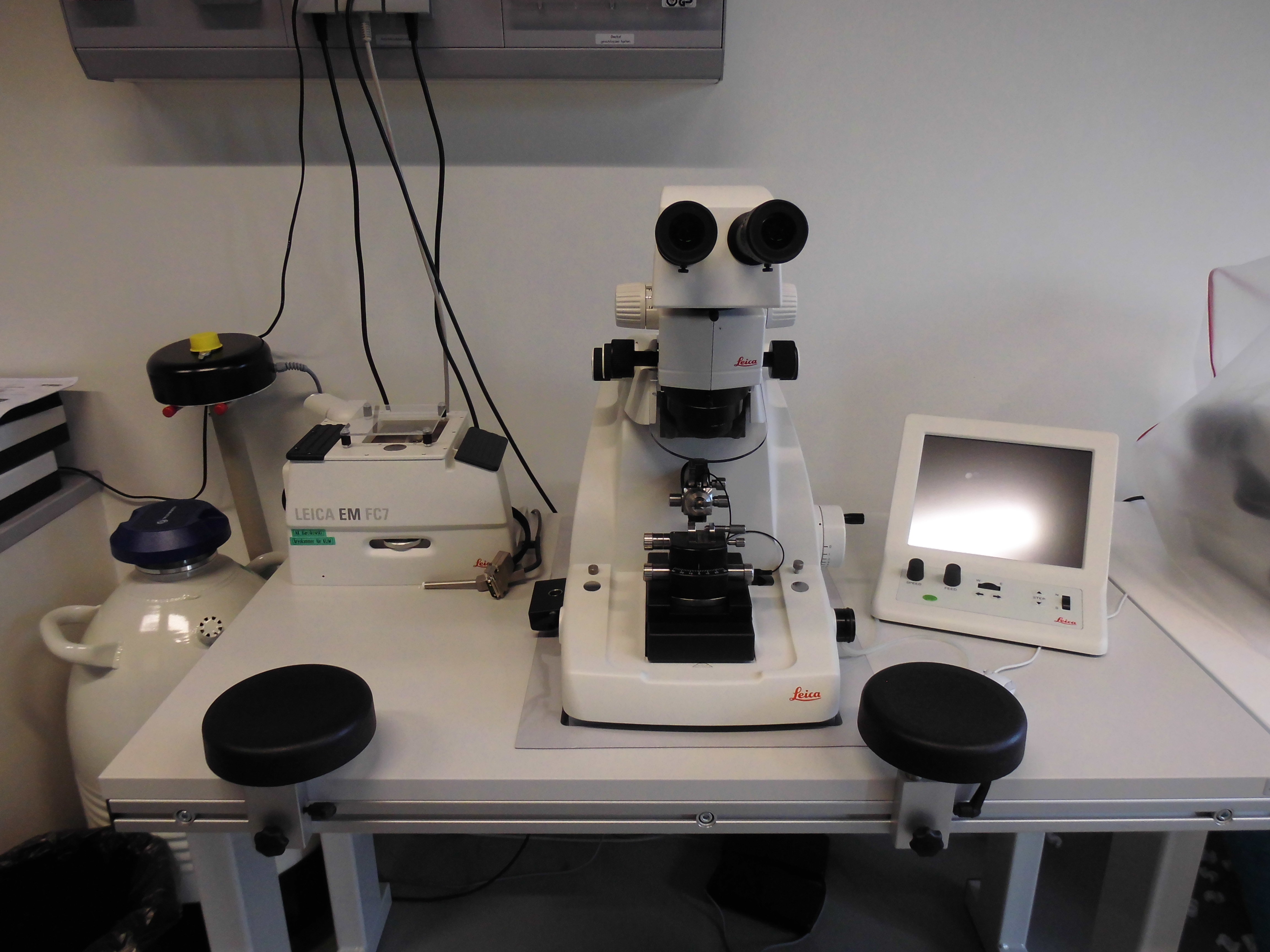
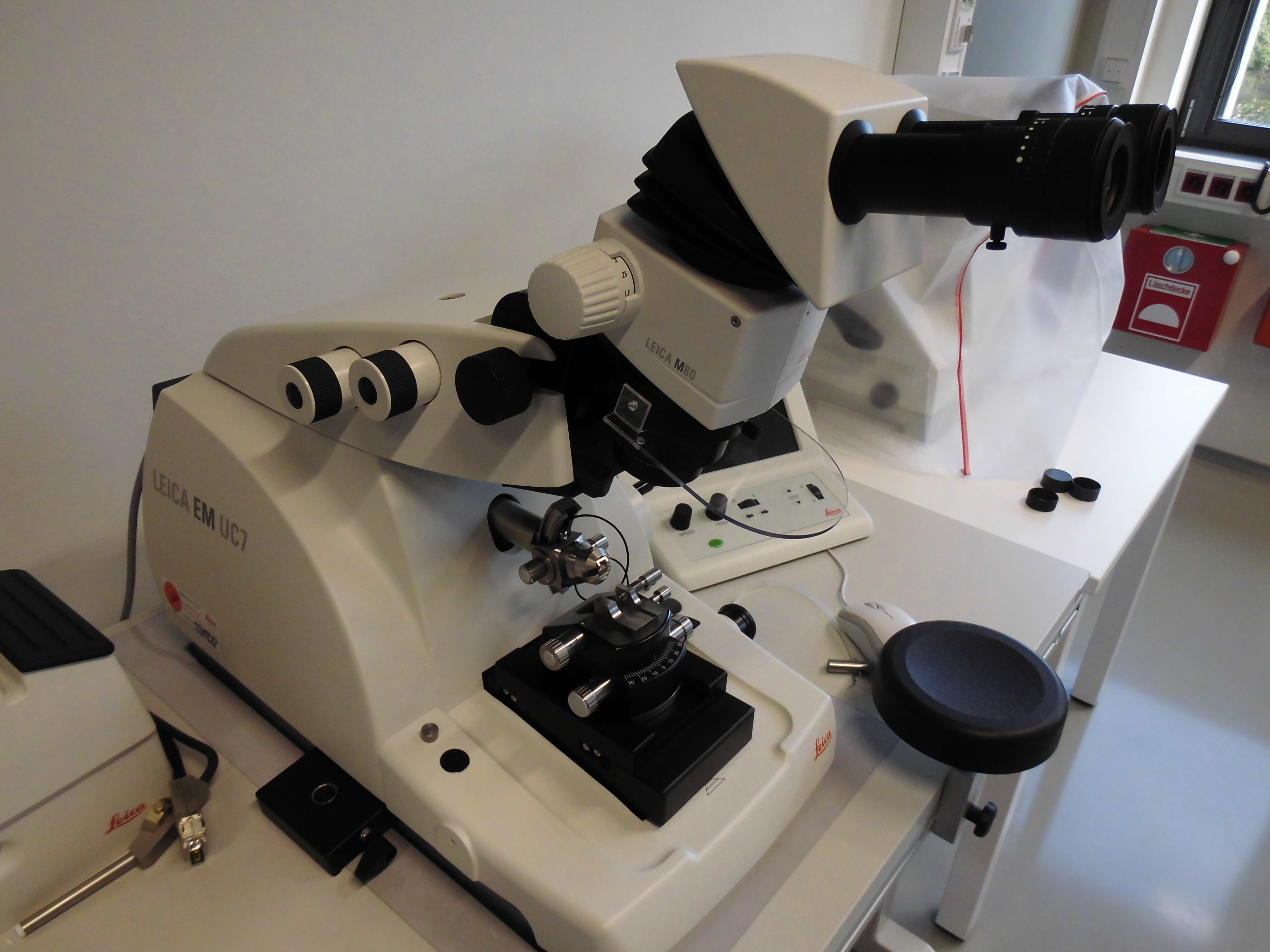
The microtome itself consists of an unmovable knife assembly and the sample holder, which can be moved. An optical microscope allows the in situ observation of the process.
The sample is being moved across the diamond knife assembly to generate the sections. After every cut the sample is moved forward by a precisely defined motion to achieve sections of equal thickness. The sections are collected in a water bath. Temperature sensitive samples can be cooled by the attachment of a cryo-chamber which allows cooling with liquid nitrogen. This is used as well, when samples are to soft to be cut. Cooling these samples hardens them and allows their sectioning.
The cutting knife has a tungsten-carbid tip with a diameter of 8 mm and rotates at 20 000 rpm with 1 µm precision. The sample can be rotated in steps of 90°.
| Magnification: | 10x – 64x |
| Light sources: | LED – adjustable brightness |
| Forward motion: | 200 µmtd |
| Segmental arch / eucentric movment : | 360° rotatable compound / +/- 22° |
| Rough knife movement N-S / O-W: | 10mm stepper/ 25 mm stepper |
| Section window: | 0,2-14 mm adjustable |
| Cutting velocity: | 0,05-100 mm/s |
| Precision: | 0,1-15 µm steps |
Cryo-chamber
| Temperature range: | +110°C to -185°C |

![]()
![]()
![]()
Cutting:
Thinning:
Polishing:
Coating:
Cleaning:
Drying: