Research
Morbidity and mortality rates after acute myocardial infarction are substantial, and the prevalence of post-infarction heart failure is increasingly relevant for health care systems. In clinical practice, limiting the extent of myocardial infarction is routinely achieved by timely and successful myocardial reperfusion. This goal is all the more important because of the inability of the human heart to regenerate lost muscle tissue. The death of cardiomyocytes provokes ventricular wall thinning, ventricular dilatation, and fibrosis, ultimately leading to left ventricular dysfunction and heart failure. Extensive loss of cardiomyocytes during ischemia and early reperfusion initiates a comprehensive, orchestrated cardiac repair process to protect functional myocardium and restore the injured areas. Therefore, the maintenance of cardiomyocyte function is the ultimate goal in myocardial infarction therapy, because cardiomyocytes form the muscles of the working heart. The repair process can be divided into the proinflammatory reaction and the resolution phase, which implies the reparative process, involving components of both the innate and the adaptive immune systems. In the early phase of reperfusion, neutrophils, monocytes, and macrophages massively invade the myocardium, while lymphocyte influx can be observed during secondary inflammatory waves. The subsequent reparative phase encompasses resolution of inflammation, (myo)fibroblast proliferation, scar formation, and neovascularization. Nevertheless, a history of myocardial infarction is associated with a 5-fold increase in the incidence of heart failure five years after infarction. It seems that the endogenous response of cardiomyocyte protection is either insufficient or crucially affected. An imbalance between the inflammatory and resolution phases toward inflammation contributes to the pathogenesis of heart failure, indicating the medical need for improving cardiac repair processes. Meeting this need requires a better understanding of the distinct cellular interfaces in reperfused acute myocardial infarction.
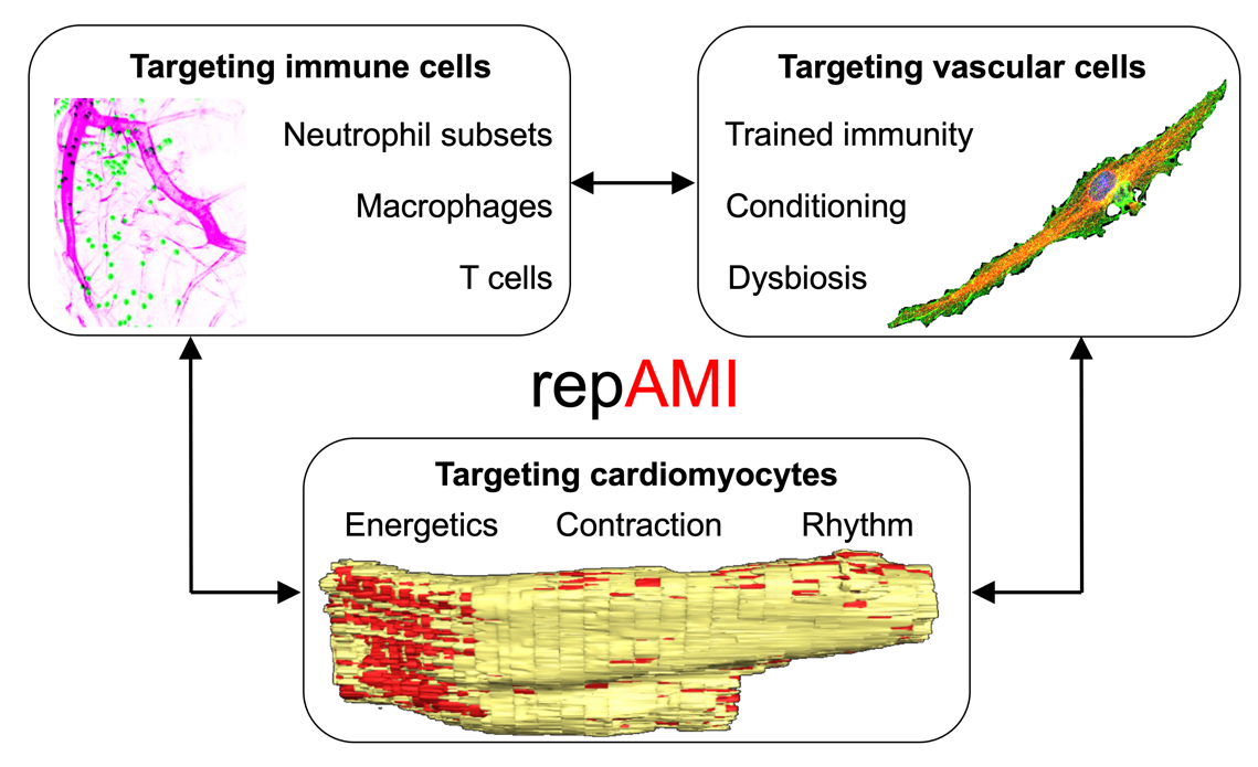
The overall research idea of the proposed RTG 2989 is the characterization of the complex interplay between specific immune cells, vascular cells, and cardiomyocytes in the processes of reperfused myocardial infarction as the basis for novel tractable therapeutic strategies. A growing body of evidence demonstrates wide phenotypic variation of innate immune cells, including neutrophils, monocytes, macrophages, and T cells in reperfused myocardial infarction. The infiltration of the immune cells into the myocardium can be related to specific regions (infarct zone, border zone, or remote zone), but the precise role of specific immune cell subsets, their spatial resolution, and their interaction with specific cardiomyocytes and vascular endothelial cells remain incompletely understood. The vascular compartment of the human heart includes at least 17 distinct cell populations, including 10 subsets of endothelial cells with arteriovenous specificities. A transcriptome profiling study of cells collected by biopsy of normal and failing hearts unmasked distinctive transcriptomic and compositional alterations in endothelial cells and their crucial role in regulating the behavior of cardiomyocytes. Excessive endothelial cell–cardiomyocyte signaling exchange may prompt a vicious cycle that causes cardiomyocyte injury and death. Furthermore, it has been suggested that endothelial cells communicate in an angiocrine crosstalk during the reparative phase.
The human heart contains a heterogenous population of cardiomyocytes in both, the left atrium and the left ventricle, each with at least five subclusters according to transcriptome profiling. The spatial localization and functions of cardiomyocyte subtypes, including cell-cell communications, remain elusive. Generally, necrotic cardiomyocytes provide the impetus for the inflammatory response. Viable cardiomyocytes in the border zone have been suggested to trigger both, inflammatory activation and inhibition in reperfused myocardial infarction. Their relative contribution to the progression and extension of postinfarction inflammation and repair remains unknown, in particular with regard to cardiomyocyte phenotypes and possible interactions with subtypes of neutrophils, macrophages, and T cells, as well as endothelial cells, as observed in recent studies on cardiomyopathy and heart failure in mouse and human tissues. Atrial cardiomyocytes are also largely compromised in reperfused myocardial infarction. It has been suggested that specific cardiac inflammation and subsequent fibrosis drive atrial cardiomyocytes into arrhythmia and atrial fibrillation. By which inter- and intracellular communications the fate of cardiomyocytes is determined in a spatiotemporal manner is incompletely understood.
Taken together, deeper insights into non-cardiomyocyte and cardiomyocyte signatures and their complex intra- and intercellular communication network may bridge current gaps and contribute to the creation of a basis for the development of effective interventions sufficient to minimize cardiac damage and dysfunction in reperfused myocardial infarction. Taking advantage of the proposed paired supervisory concept of a clinical and a basic science expert, the proposed RTG 2989 will systematically investigate (i) the plasticity and control mechanisms of immune cells, (ii) the involvement of the vascular endothelium, and (iii) the heterogeneous functionality, plasticity, and remodeling of cardiomyocytes in repAMI in three distinct research areas and 11 projects supervised by a Tandem PI team.