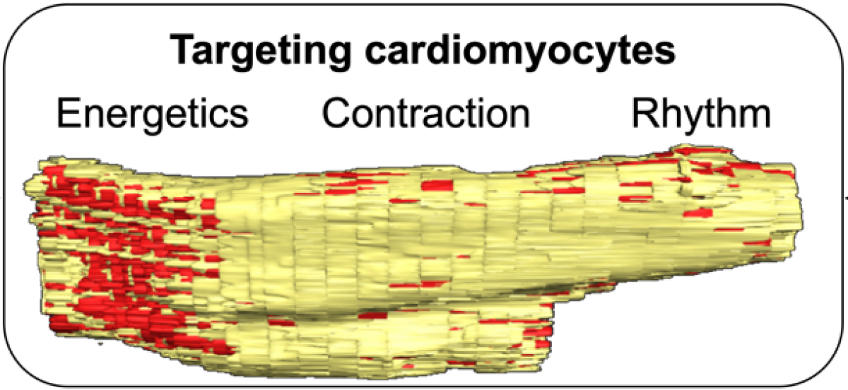
Research Area 3: targeting cardiomyocytes
The explosive burst of cardiomyocyte death in early reperfused myocardial infarction and the subsequent maladaptive remodeling of surviving cardiomyocytes with changes in Ca2+ cycling and aggravation of inflammatory activities result in deterioration of cardiac output. This scenario inflicts a secondary stress on the heart in the form of increased afterload that is chronic and unremitting, providing a strong stimulus for cardiomyocyte death, which initiates a vicious cycle of unfavorable geometrical remodeling that stimulates yet more cell death and progressing heart failure. Members of the BCL-2 family are suggested to be important players in the development of pressure-overload HF exerting adverse effects by inducing mitochondrial Ca2+ overload, apoptosis and left ventricular interstitial fibrosis. In addition, cardiomyocytes are strongly involved in the two-way communication with other non-resident and resident cells detectable in reperfused myocardial infarction, particularly immune and endothelial cells and fibroblasts. Whether and how cardiomyocyte heterogeneity and its spatiotemporal functional dynamics, including cell-cell-communications, determine the outcome remains elusive. Disintegration of atrial cardiomyocyte homeostasis often results in substantial arrhythmia followed by atrial fibrillation as the most frequent supraventricular rhythm disorder, with an estimated incidence of up to 21%. It remains unclear, however, how myocardial infarction, mainly located in the left ventricle, drives atrial cardiomyocytes into atrial fibrillation. The reason for new-onset atrial fibrillation is multifactorial and is linked to cardiac inflammation and consequential interstitial fibrosis in the early phase of reperfused myocardial infarction. The composition of atrial immunologic infiltrates is not well known, and their effect on the morphology and functionality of atrial cardiomyocytes remains to be determined. There has been cumulative evidence for a critical role of NLRP3 inflammasome signaling in patients with common atrial fibrillation forms and those predisposed to post-operative atrial fibrillation. Interestingly, NLRP3 inflammasome activation is linked to increased susceptibility also in mice with high-fatty diet, suggesting a potential causal relationship between atherosclerosis and atrial fibrillation. The precise immune response pattern in atria and the functional and morphological consequences remain to be determined. While cardiomyocytes may respond to myocardial infarction with significant morphological and functional adaptions, heart failure remains one of the highly relevant long-term sequelae for patients.
Recent studies imply the plasticity and substantial recovery potential of human cardiac cells, even in end-stage heart failure. Transcriptomes of cardiomyocytes, endothelial cells, and fibroblasts from patients with heart failure revealed contraction and metabolism as the crucial factors, which were impaired after worsening of heart function. Implantation of an left ventricular assist device (LVAD) is often the last therapeutic option for patients with terminal heart failure and is the most common form of mechanically assisted circulatory support. Indications for LVAD include “bridge to recovery”, “bridge to transplant” or “destination therapy”. Whereas the last two indications have been well studied, “bridge to recovery” remains a challenge. Some LVAD patients demonstrate myocardial recovery enabling explantation of the device without further surgical treatment such as re-implantation of a VAD or heart transplantation. Although several molecular changes, such as change in Ca2+ handling proteins, in cytoskeletal pathways, in collagen and matrix protein composition, and in the immune system have been identified as a result of chronic left ventricular unloading, the clinical importance and correlation remain unclear. It is likely that many other protein changes occur that are unknown; their discovery will require unbiased screening of patient samples. Further insights into the mechanism of cell-state reversal may lead to better exploitation of innate recovery processes of the human heart. Heart transplantation is the definitive solution for failing hearts. However, donor hearts are challenged with phases of hypoxia/ischemia and reperfusion in the re-implantation procedure. Improved preservation and reconditioning of machine-perfused donor hearts is essential for increasing retrieval and the acceptance rate of donor hearts. Machine perfusion can preserve the donated heart in a beating, semi-physiologic dynamic status. Recent findings during the past decade provided increasing evidence that machine perfusion may be considered superior to conventional cold storage on ice. The maintenance of an aerobic metabolism seems to be more protective, especially now that ischemic times are longer, because most of the transplant patients are undergoing mechanically assisted therapy. A dynamic platform in machine perfusion therapy allows for standardized preservation and reconditioning therapies. Limitations in therapy today are the availability of enough donor blood to run the system and the fact, that longer machine perfusion times may lead to the formation of tissue edema due to extravasation of perfusion solution (because of structural degeneration of the endothelial cells/glycocalyx) in the long run. Cardiomyocytes possess an abundant and complex mitochondrial network. In reperfused myocardial infarction, mitochondrial damage drives alterations in metabolism, a decline in bioenergetic capacity, inflammatory responses, and impaired mitophagy in the remote zone, which compromises cardiomyocyte integrity and cardiac output. Recent evidence suggests that cardiomyocytes clear mitochondrial debris via membranous vesicles, with substantial interplay between intracellular mitophagy and tissue-resident macrophages. How this multiplicity of processes is regulated and altered in reperfused myocardial infarction at the level of the mitochondrial network is still unclear. The specific aims for this research area are (i) characterization of cardiomyocyte communication network, plasticity and fate landscape in the peri-infarct and remote myocardium, (ii) evaluation of inflammation-induced atrial remodeling, (iii) unbiased screening for molecular targets from ventricular unloading, (iv) preservation of donor hearts through novel oxygen carriers, and (v) identifying how mitochondrial remodeling and clearance shape cell fate decisions. The projects will be highly interactive and will involve experimental and clinical approaches.