Schwarzer Lab – Research Focus
Schwarzer Lab · Research Focus

FROM HOST-CELL STATE TO THERAPEUTIC OPPORTUNITY

Learn more about our research areas and projects

How we work

Model relevant biology

We combine cell-line systems, primary human cells, tissue-derived material, and clinical samples to study viral infection in contexts that reflect disease-relevant biology.

Measure cell states deeply

We use flow cytometry, infection assays, transcriptomics, single-cell approaches, metabolic profiling, imaging, and perturbation screens to capture how infected and exposed cells change.

Translate mechanisms

We use mechanistic insight to nominate antiviral targets, biomarkers, host-directed strategies, and experimental workflows that can support translational virology.

HIV reservoirs and cure research
HIV reservoir and cure research overview
HIV · Latency · Cure strategies

Understanding and targeting the latent HIV reservoir

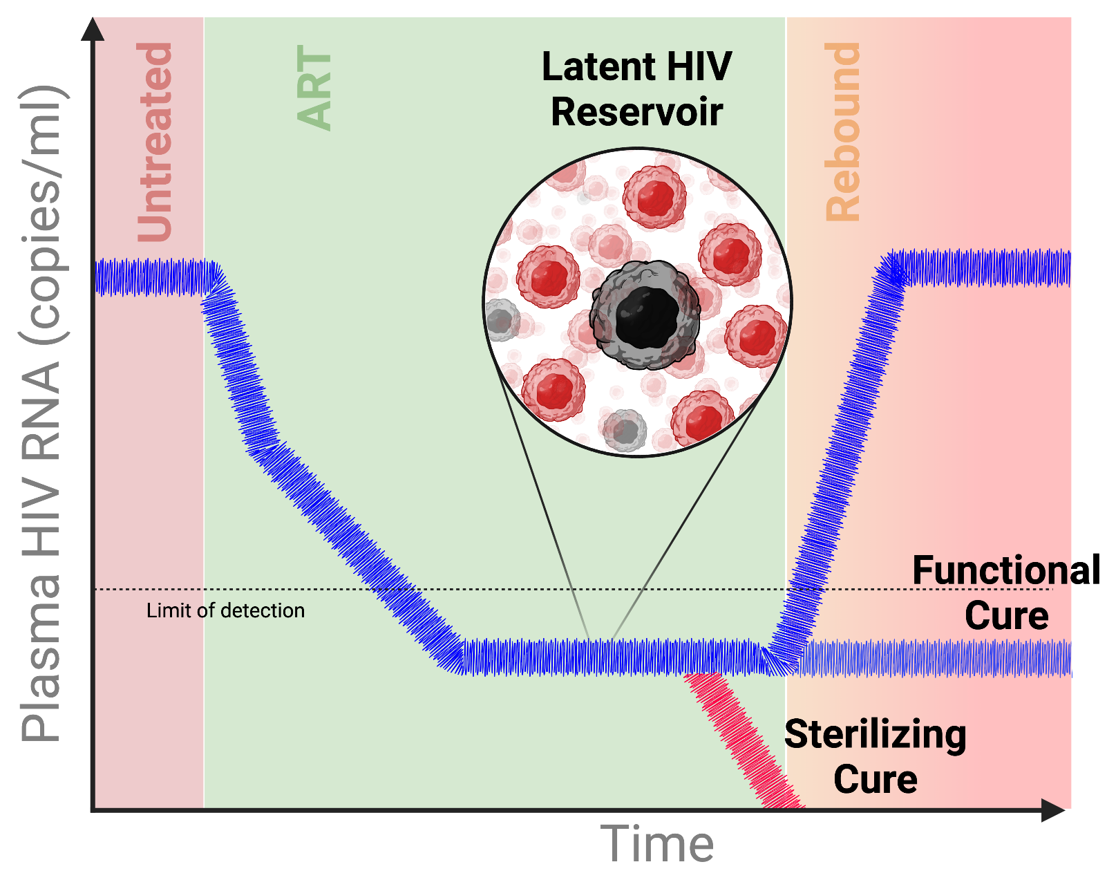
Antiretroviral therapy has transformed HIV infection from a fatal disease into a manageable chronic condition. However, ART does not eliminate latently infected cells. These cells can remain dormant for years or even decades and can reignite infection if therapy is interrupted.

Our central goal is to understand what latent reservoir cells are, where they reside, how they are maintained, and how they can be therapeutically targeted. We study reservoir seeding, survival, and reactivation from latency, while also considering the broader mechanisms of HIV pathogenesis and disease progression.

We use high-dimensional systems virology, CRISPR-based perturbation, latency models, primary cell systems, and samples from people living with HIV to dissect reservoir biology at mechanistic depth.

Research statement

We view HIV cure strategies as necessarily multifactorial. Durable remission will likely require reservoir reduction, control of residual latent proviruses, and immunological interventions that prevent rebound and disease progression.

Models Cell-line latency systems, primary cells, and clinical samples.
Readouts Reservoir size, transcriptional activity, reactivation, and cell state.
Tools CRISPR, systems virology, single-cell and high-dimensional profiling.
HIV pathogenesis
HIV infection outcome and pyroptosis schematic
Inflammation · Pyroptosis · Tissue CD4 T cells

Inflammatory circuits that drive HIV disease progression

HIV pathogenesis is strongly shaped by incomplete, abortive infections in tissue-resident immune cells. In affected CD4 T cells, intracellular recognition of viral material can trigger inflammasome activation and caspase-1-dependent pyroptosis.

This inflammatory cell death contributes to a vicious cycle: dying cells amplify local inflammation, recruit new HIV-susceptible cells, and thereby create conditions for further abortive infection and immune-cell loss.

We study how extrinsic factors, host-cell context, and tissue environments modulate pyroptotic cell death. A better understanding of these determinants could inform targeted anti-inflammatory strategies, personalized therapeutic approaches, or diagnostic tools.

Research statement

HIV-induced pyroptotic cell death is best captured in tissue-derived CD4 T cells. We therefore work with CD4 T cells from tonsillar tissue and combine flow cytometry, plate-reader assays, and complementary model systems to quantify inflammatory cell death after in vitro HIV infection.

Tissue context Tonsillar CD4 T cells from clinical material.
Assays Flow cytometry and plate-reader-based pyroptosis readouts.
Translation Cell-line models for scalable testing of anti-inflammatory interventions.
Antiviral discovery
Antiviral research composite showing nanoparticles, proteins, and dose-response assays
Therapeutics · Resistance · Delivery

Developing and characterizing antiviral strategies

Recent outbreaks and pandemic threats have made clear that better antiviral strategies are still urgently needed. Classical treatments often fail to fully control infection at the level of individual patients or larger populations.

Our lab contributes to antiviral discovery through interdisciplinary collaborations. We investigate virus-specific and pan-antiviral approaches, mechanisms of drug resistance and evasion, and host-dependent therapy failure.

Our work spans multiple viral families and includes antiviral proteins, nanoparticle and nanofiber delivery systems, natural compounds, and mechanistic studies of antiviral activity.

Research statement

We have contributed to work on bispecific SARS-CoV-2 antibodies, improved nanoparticle formulations of antiretroviral drugs, and the pan-antiviral activity of the bovine seminal plasma protein PDC-109. We continue to dissect these mechanisms and test improved delivery architectures with national and international collaborators.

Discovery Collaborative screening of antiviral candidates and natural compounds.
Delivery Nanoparticle and nanofiber formulations for antiretroviral applications.
Mechanisms Resistance, evasion, host-dependence, and pan-antiviral activity.
Immunometabolic virology
Immunometabolic virology overview
Metabolism · Microenvironment · Omics

How metabolic state shapes viral infection and immunity

Viral infections and cellular metabolism are deeply interconnected. Viruses such as CMV, HCV, and HIV can exploit glycolysis, oxidative phosphorylation, and mitochondrial function to redirect cellular resources. At the same time, the metabolic state of the host cell can influence viral entry, replication, and egress.

Infection biology is also shaped by external factors such as oxygenation, temperature, paracrine signaling, local tissue microenvironments, sex, and age. We are particularly interested in how these contextual variables reshape immune regulation and viral behavior.

We use transcriptomics and other high-dimensional discovery approaches to study viral infections under defined microenvironmental conditions. Seahorse assays, mass spectrometry, CRISPR technologies, and pharmacological tools help us identify metabolic dependencies and potential intervention points.

Research statement

We view metabolism as a crucial determinant of viral replication and infection outcome. Understanding how metabolic and immunologic features are shaped by host context and microenvironment may reveal new therapeutic and diagnostic opportunities.

Discovery Transcriptomics and high-dimensional profiling under defined conditions.
Metabolic assays Seahorse, mass spectrometry, and pathway-focused readouts.
Perturbation CRISPR and pharmacological tools to test metabolic dependencies.
Emerging virus and orthohantavirus research
Orthohantavirus project pipeline and microscopy panels
Orthohantaviruses · Cell biology · Host-directed antivirals

Host–virus interfaces in emerging RNA virus infections

Emerging viruses are pathogens that newly appear in a population or show rapidly increasing incidence or geographical spread. Orthohantaviruses are zoonotic, enveloped RNA viruses with small mammals such as rodents, shrews, and bats as natural hosts. Human infection typically occurs through inhalation of contaminated excreta.

Orthohantaviruses contain a tri-segmented negative-sense RNA genome encoding a compact set of viral proteins: the glycoproteins Gn and Gc, the nucleoprotein N, the RNA-dependent RNA polymerase, and in some cases an accessory NSs protein.

We investigate the intracellular dynamics and host interactions of orthohantavirus proteins, with particular focus on glycoprotein entry functions, nucleoprotein biology, cytoskeletal remodeling, and interactions with P-bodies.

Research statement

Our goal is to understand the host factors that are critically involved in the hantavirus life cycle. We use interactomics, CRISPR technologies, and high-resolution quantitative microscopy to identify host–virus interfaces that may inform host-directed antiviral strategies or diagnostic markers.

Virus biology Entry, replication, protein dynamics, and viral assembly interfaces.
Cell biology Cytoskeleton remodeling, P-body interactions, and infected-cell organization.
Technologies Interactomics, CRISPR perturbation, and quantitative microscopy.