AG Schleberger Lab equipment
2D Materials Lab
We have various equipment and methods at our disposal for the production and characterization of 2D materials. These include several tube furnaces for chemical vapor deposition, with which we mainly produce TMDCs (MoS2, WS2, MoSe2, WSe2, SnS2). We also have a glovebox for controlled environmental conditions, a stacker for the production of heterostructures, optical and confocal Raman/photoluminescence and scanning probe microscopes. Raman and PL can be performed under non-ambient conditions and at variable temperatures in conjunction with electrical measurements. Electrical devices are usually processed by us using maskless UV lithography in the AG Lorke clean room. For more advanced requirements, we can also use electron beam lithography. Finally, we have various ion sources and a plasma etcher at our disposal for the controlled modification of our materials.
In addition, the Schleberger group operates a variety of UHV-setups with in situ surface preparation techniques such as heating, sputtering, ovens for epitaxy and deposition of molecules.
Raman/PL-microscope:
- WiTec Raman/PL/AFM with 457 nm, 532 nm, and 633 nm lasers & Linkam stage
- Renishaw Raman/PL/AFM with 532 nm and 638 nm lasers with fiber-optics for in-situ analysis
- Custom-build Raman-vacuum stage
Scanning probe microscopes:
- Veeco Dimension 3100 (ambient conditions, together with AG Horn von Hoegen)
- Omicron RT-STM/AFM UHV with RHK SPM-1000 & PLL Pro2
- Omicron VT-STM/AFM UHV with Matrix controller
- RHK VT-AFM UHV 750 with R9 controller and optical access for micron-sized samples
- WiTec AFM head in connection with Ramanmicroscope
Spectroscopy:
- MCP LEED and standard LEED
- Spectra and Phi X-ray sources
- Staib DESA-100 and PHI 500 electron spectrometer for AES and XPS
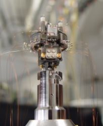
Beamlines for highly charged ions:
- UHV-setup equipped with Dresden EBIS ion source for the production of highly charged ions up to Epot = 66 keV, e.g. Xe46+, charge state separation, deccelaration stage (1 keV*q), ToF-analysis and VUV-laser for post-ionization
- Dresden EBIT ion source for the production of highly charged ions up to Epot = 45 keV, e.g. Xe42+ mounted to a UHV scanning probe microscope for in-situ analysis of HCI induced defects
Experiments with swift heavy ions are conducted at the
- GANIL/CIMAP in France (IRRSUD, SME, ARIBE) in collaboration with H. Lebius
- GSI in Darmstadt (permanent STM/AFM setup at the M-branch)
- RBI in Croatia in collaboration with M. Karlusic

HICS setup consisting of 1: EBIT, 2: Extraction system, 3: Magnet, 6: Irradiation chamber