SHERPA
SHERPA: an image analysis tool for diatomsShape Recognition, Processing and Analysis
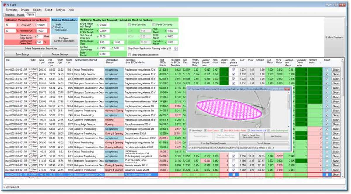
SHERPA is a software tool developed by Michael Kloster implementing a highly customizable image processing workflow for the identification and quantification of object outlines. SHERPA was mainly developed for the analysis of bright field microscopic images of diatom valves, but might be useful for other applications as well.
When using SHERPA for your research, please cite our publication describing it:
Kloster M., Kauer G. & Beszteri B. (2014): SHERPA: an image segmentation and outline feature extraction tool for diatoms and other objects. BMC Bioinformatics 15:218.
SHERPA is provided for your use for free but without any guarantees, please read the below disclaimer before downloading.
THE SOFTWARE AVAILABLE HERE IS PROVIDED ''AS IS'' AND ANY EXPRESSED OR IMPLIED WARRANTIES, INCLUDING, BUT NOT LIMITED TO, THE IMPLIED WARRANTIES OF MERCHANTABILITY AND FITNESS FOR A PARTICULAR PURPOSE ARE DISCLAIMED. IN NO EVENT SHALL AWI OR ITS STAFF BE LIABLE FOR ANY DIRECT, INDIRECT, INCIDENTAL, SPECIAL, EXEMPLARY, OR CONSEQUENTIAL DAMAGES (INCLUDING, BUT NOT LIMITED TO, PROCUREMENT OF SUBSTITUTE GOODS OR SERVICES; LOSS OF USE, DATA, OR PROFITS; OR BUSINESS INTERRUPTION) HOWEVER CAUSED AND ON ANY THEORY OF LIABILITY, WHETHER IN CONTRACT, STRICT LIABILITY, OR TORT INCLUDING NEGLIGENCE OR OTHERWISE ARISING IN ANY WAY OUT OF THE USE OF THIS SOFTWARE, EVEN IF ADVISED OF THE POSSIBILITY OF SUCH DAMAGE.
Click here to download the latest version, SHERPA 1.1c.
An older version is still available on our old SHERPA page.
Requirements:
Microsoft .NET Framework 4.62, the Microsoft Visual C++ 2010 SP1 Redistributable Package (x64) and the Microsoft Visual C++ Redistributable for Visual Studio 2012 Update 4 (x64) (please download file "VSU_4\vcredist_x64.exe") are required for running SHERPA 1.1c (if not already present on your system).
Video Digitaler Workflow
Dies ist eine (sehr) kurze Einführung in unseren digitalen Slidescanning und Annotations-Workflow für Diatomeen.
Datenschutz-Hinweis
Mit Abruf dieses Videos wird Ihre IP-Adresse an den externen Anbieter des Videos übermittelt. Dieser kann auch Cookies auf Ihrem Gerät setzen.
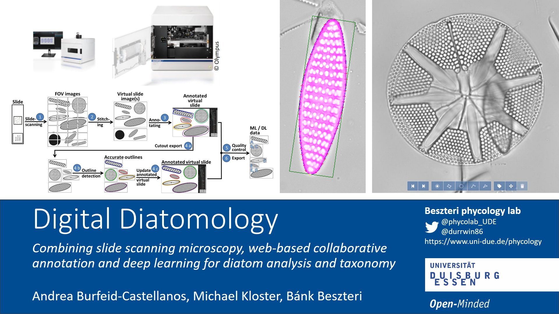
Digital Diatomology: Combining slide scanning microscopy, web-based collaborative annotation, and deep learning for diatom analysis
Andrea Burfeid Castellanos und Michael Kloster stellen digitale Varianten der Lichtmikroskopie für die Diatomeenanalyse vor im Rahmen der Diatom Web Academy am 17.11.2020.
Introduction To Annotating Diatom Virtual Slides With Biigle
This screencast by Michael Kloster introduces the basic functionality of the BIIGLE 2.0 platform we use for web-based collaborative taxonomic annotation of high resolution diatom slide scans (virtual slides). For another account on this methodology, as applied for teaching diatom taxonomy during lockdown, see this post on the Young Diatomists Blog by Andrea Burfeid!
ProtocolDigital diatom analysis workflow
In den folgenden Veröffentlichungen beschreiben wir, wie wir hochauflösendes Slide-Scanning in Kombination mit web-basierter taxonomischer Annotierung und Deep Learning für die Diatomeen-Analyse einsetzen:
Earlier, using SHERPA and a special slide scanning microscope, we proposed an implementation of the two-step digital diatom analysis workflow originally drafted by ADIAC. You can find a detailed description here:
Kloster M., Esper O., Kauer G. & Beszteri B. (2017):Large-Scale Permanent Slide Imaging and Image Analysis for Diatom Morphometrics. Applied Sciences 7(4): 330
Use casesOur publications using SHERPA
Burfeid-Castellanos, A.M., Kloster, M., Cambra, J., Beszteri, B. (2020). Both hydrology and physicochemistry influence diatom morphometry. Diatom Research 35: 315-326
Postel, U., Glemser, B., Salazar Alekseyeva, K., Eggers, S.L., Groth, M., Glöckner, G., John, U., Mock, T., Klemm, K., Valentin, K., Beszteri, B. (2020). Adaptive divergence across Southern Ocean gradients in the pelagic diatom Fragilariopsis kerguelensis. Molecular Ecology, doi: 10.1111/mec.15554.
Glemser, B., Kloster, M., Esper, O., Eggers, S.L., Kauer, G. and Beszteri, B. (2019). Biogeographic differentiation between two morphotypes of the Southern Ocean diatom Fragilariopsis kerguelensis. Polar Biology 42: 1369-1376.
Kloster, M., Rigual Hernández, A.S., Armand, L.K., Kauer, G., Trull, T.W. and Beszteri, B. (2019). Temporal changes in size distributions of the Southern Ocean diatom Fragilariopsis kerguelensis through high-throughput microscopy of sediment trap samples. Diatom Research, on line early.
Kloster, M., Kauer, G., Esper, O., Fuchs, N. and Beszteri, B. (2018). Morphometry of the diatom Fragilariopsis kerguelensis from Southern Ocean sediment: High-throughput measurements show second morphotype occurring during glacials. Marine Micropaleontology 143: 70-79.
Beszteri, B., Allen, C., Almandoz, G.O., Armand, L., Barcena, M. A., Cantzler, H., Crosta, X., Esper, O., Jordan, R. W., Kauer, G., Klaas, C., Kloster, M., Leventer, A., Pike, J. and Rigual Hernández, A.S. (2018). Quantitative comparison of taxa and taxon concepts in the diatom genus Fragilariopsis: a case study on using slide scanning, multiexpert image annotation, and image analysis in taxonomy. Journal of Phycology 54: 703-719.
Data and analysis scripts
Example images for testing SHERPA: a typical sample of Fragilariopsis kerguelensis valves from sediment core PS1768-8 (the slide was kindly provided to us by Rainer Gersonde). The original high resolution TIFs are available from PANGAEA.
R code for Sellaphora analyses of images from Mann et al. (2004)
Our data sets and R code accompanying the analyses in our above listed papers are on PANGAEA.
Data from publication "Deep learning-based diatom taxonomy on virtual slides"
Supplement III: Data and R scripts
Supplement IV: Detailed results for VGG16_1FC
(see the paper here)