Technical background
InformationUsed technologies
UDE BioSLiDES uses a number of different technologies and partly in-house developed software for the digitization and visualization of microscopic specimens:
-
The imaging process
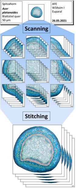
Image acquisition: The specimens are scanned using an Olympus VS200 slide scanner with different high-resolution objectives (20×/0.80, 40×/1.40 or 60×/1.42). The device scans the entire surface of the specimen in the form of slightly overlapping fields of view, whereby approx. 40-80 different focus positions are imaged at each position. The distance between adjacent focal planes usually corresponds to half the depth of field of the objective, occasionally to the full depth of field, i.e. every detail of the specimen is in focus in at least one focal plane. For each individual focal plane, the up to several thousand individual field of view images are then combined by a process called stitching into a single large image. Depending on the specimen, this comprises up to several thousand megapixels. The entire process is illustrated in the figure on the right.
- Post-processing: The proprietary file format of the slide scanner must be translated into suitable data formats and processed further for use in UDE BioSLiDES. A particular sticking point here is the size of the individual focal planes, as these can be larger than standard image file formats such as JPEG or TIFF allow for. In order to facilitate the reuse of the image data outside of UDE BioSLiDES, the various planes are additionally combined by means of focus stacking to form an extended focus image, which sharply depicts the examination object over its entire depth in a single image plane. Additional information about the specimen and its preparation, as well as annotations must be entered and integrated manually. A number of open source, commercial and self-developed software tools as well as open file formats such as JPG, (Big)TIFF and JSON are used for the individual processing steps.
- Visualization: The specimen viewer was developed specifically for UDE BioSLiDES in JavaScript/HTML and is based on a number of open source tools and frameworks; basis for the visualization is OpenSeadragon , which we extended to include the functions for focusing through the specimen. In addition, the viewer integrates the display of text-based information and annotations (the latter implemented via Annotorious ) and allows to interact with the digital specimen in a variety of ways.
- Sticking points: UDE BioSLiDES required the development of various different software tools, but the main problem is handling the gigantic amounts of data that are generated during the production of the digital specimens. The currently most extensive specimen comprises 236 gigapixels (over 250 billion pixels) in the form of 90,000 individual files with a total size of approx. 32 GB; all 200 specimens together comprise approx. 1.6 TB distributed over more than 2,000,000 files.