EVOS™ M7000
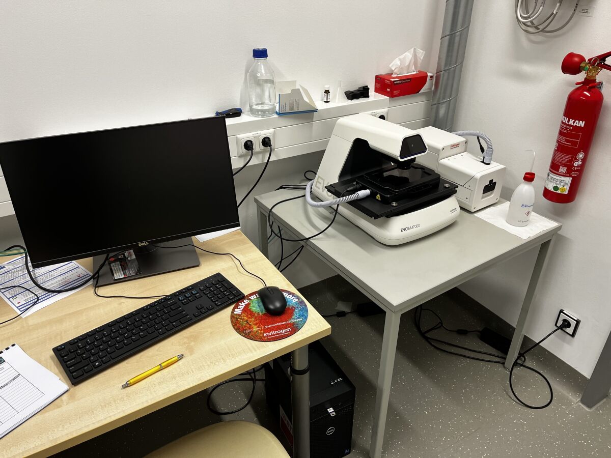
Widefield Systems EVOS M7000 Imaging System
Room: T03 R01 D63
Main Applications
- fully automated multi-channel fluorescence imaging
- transmitted light applications (brightfield, phase contrast) and color brightfield
- multi-dimensional live cell + fixed specimen imaging
- multi-position acquisition, area scan with monatge or tile-stitching, Z-Stacking
- 2D/3D deconvolution
Technical Specifications
Microscope and Accessories
- Inverted microscope stand
- software based autofocus (fluorescence or transmitted light)
- On-Stage Incubator environmental chamber with temperature & CO2 control for live cell imaging
Cameras
- High-sensitivity 3.2 MP monochrome CMOS camera (2048 x 1536 pixels) with 3.45 μm pixel resolution
- High-sensitivity 3.2 MP color CMOS camera (2048 x 1536 pixels) with 3.45 μm pixel resolution
Light sources
adjustable-intensity LED light cubes:
| name |
Excitation |
Emission |
| DAPI | 357/44 nm | 447/60 nm |
| CFP (upon request) | 445/45 nm | 510/42 nm |
| GFP | 482/25 nm | 524/24 nm |
| YFP (upon request) | 500/24 nm | 542/27 nm |
| RFP | 531/40 nm | 593/40 nm |
| TexasRed | 585/29 nm | 628/32 nm |
| Cy5 | 628/40 nm | 692/40 nm |
Objectives
| name |
magnification |
NA | immersion |
| EVOS FL 10x LWDPH | 10x | 0.30 | dry |
| EVOS FL 20x LWDPH | 20x | 0.45 | dry |
| Olympus XApo 20x | 20x | 0.8 | dry |
| Olympus UPlanFL 60x CC | 60x | 0.90 | dry |
| Olympus XApo 60x | 60x | 1.42 | oil |
Software
EVOS M7000 acquisition software and Celleste 7 Image Analysis Software including 2D- and 3D-Devonvolution module