Special Equipment
Confocal Laser Scanning Microscopy (CLSM)
Confocal Principle
The big advantage of confocal microscopy is the possibility to collect light exclusively from a single plane.
A pinhole sitting conjugated to the focal plane (i.e.confocal) keeps light from the detector that is reflected/emitted from others than the focal plane.
The laser scanning microscope scans the sample sequentially point by point and line by line and assembles the pixel information to one image. That way optical slices of the specimen are imaged with high contrast and high resolution in x, y and z.
By moving the focus plane single images (optical slices) can be put together to build up a three dimensional stack that can be digitally processed afterwards.
More details can be found on the Website of Zeiss
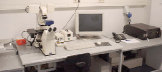
On Site Instrumentation
Laser scanning modul LSM 510 from Zeiss mounted on an inverted microscope stage Axiovert 100 M BP. The system is equipped with an argon laser (possible excitation wavelenght: 458 nm, 488 nm and 514 nm; 25 mW), to helium neon lasern (possible excitation wavelenght: 543 nm; 1 mW and 633 nm; 5 mW) and a argon ionen UV laser (possible excitation wavelenght: 351 and 364 nm; 80 mW).
The following lenses are available: Plan-Neofluar 10x / 0.30, Plan-Neofluar 20x / 0.50, Plan-Neofluar multi immersion 25x / 0.8 imm. corr., Plan-Neofluar 40x / 1.30 oil, C-Apochromat 40x / 1.20 water corr., LD Achroplan 40x / 0.6 corr., LD Achroplan 63x / 0.75 corr., Plan-Apochromat 63x /1.40 oil DIC and Plan-Neofluar 100x / 1.30 oil.
The sytem is controled by a PC with the LSM 510 software (release 3.2, Zeiss). Image processing and 3D visualizations are possible with the AxioVision software (version 3.1, Zeiss Vision), 3DforLSM (Zeiss) and ImageVisArt (Zeiss). Quantitative image analysis can be done with the KS 300 software package (version 3.0 SP9, Zeiss Vision). Additionaly, the system is equipped with the cooled high resolution CCD camera AxioCam HRc (Zeiss).