Videos
Angular Orbital Momentum
Transmitting huge volumes of data and even moving matter using light: these are two of the visions that physicists working in the field of plasmonics have for the future. In a collaboration with colleagues from Haifa (Israel), Kaiserslautern and Stuttgart, Scientists at the Center for Nanointegration (CENIDE) at the University of Duisburg-Essen succeeded in producing nanometer-sized plasmon swirls on a metal surface, filming the cycles at 100 attosecond intervals.
Source: Spektor et al., SCIENCE 355 (2017) p. 1187
The complete press release can be found here.
Datenschutz-Hinweis
Mit Abruf dieses Videos wird Ihre IP-Adresse an den externen Anbieter des Videos übermittelt.
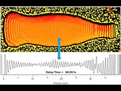
Electron Waves Running Across Microscopic Silver Bottle
Source: Microscopy team around Prof. Dr. Frank Meyer zu Heringdorf,
University of Duisburg-Essen
Datenschutz-Hinweis
Mit Abruf dieses Videos wird Ihre IP-Adresse an den externen Anbieter des Videos übermittelt.
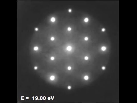
LEEM Tutorial: Pb sqrt-3xsqrt3 LEED Pattern
Imaging of LEED diffraction patterns using a LEEM is special, as the k-space scaling is independent of the electron energy - in contrast to a regular (rear-view) LEED, where the spots move on the screen when the electron energy is changed. The example shows a sequence of LEED patterns, recorded with LEEM, of a Pb submonolayer on Si(111) at different electron energies. The starting energy is 0 eV, i.e., the diameter of the Ewald Sphere is zero. As the electron energy is increased, more spots appear as the Ewald Sphere gets larger. The positions of the spots, however, do not change.
Source: Microscopy team around Prof. Dr. Frank Meyer zu Heringdorf,
University of Duisburg-Essen
Datenschutz-Hinweis
Mit Abruf dieses Videos wird Ihre IP-Adresse an den externen Anbieter des Videos übermittelt.
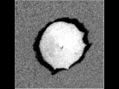
Ag Desorption from Si(111)
This PEEM movie shows the decay of an Ag island on Si(111). The interesting contrast ist caused by the particular decay mechanism: Ag desorbs from the surface, and the island in the center feeds Ag atoms into the surface layer as it decays. This mechanism creates a coverage gradient on the surface. As the coverage drops below a certain critical coverage, the surface reconstruction changes locally, which causes the strong contrast in the movie. At the end of the movie the island starts thermomigration, taking its diffusion field with it.
Source: Microscopy team around Prof. Dr. Frank Meyer zu Heringdorf,
University of Duisburg-Essen
Datenschutz-Hinweis
Mit Abruf dieses Videos wird Ihre IP-Adresse an den externen Anbieter des Videos übermittelt.
Silicon Desorption from a Si(001) Surface
This is a dark-field Low Energy Electron Microscope (LEEM) movie of a Silicon (001) surface. The imaging conditions are such that the black and white areas in the movie resemble atomically flat terraces. Between bright and dark regions is a 0.136 nm high atomic step. We are imaging at high temperatures (around 1000°C). The rapid movement of the boundary between bright and dark regions resembles thermal fluctuation of the atomic steps. Also, the temperature is sufficiently high for Silicon to evaporate from the surface. In the movie this is visible as the overall movement of the boundary between bright and dark regions. The hill in the center of the image evaporates in a atomic layer-by-layer fashion (the brightness changes periodically between bright and dark), until it disappears towards the end of the movie. At this point the step-stiffness (a rubber-band-like property of the steps) pulls the steps straight. Imaging of Si(001) like this is standard in LEEM, but this movie at high temperatures is a particularly nice version. Note: the movie is corrected for sample drift. The black edge visible at the lower right in frames 2500 and higher is the edge of the Imaging detector.
Source: Microscopy team around Prof. Dr. Frank Meyer zu Heringdorf,
University of Duisburg-Essen
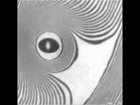
Standing Wave Formation
Source: Microscopy team around Prof. Dr. Frank Meyer zu Heringdorf,
University of Duisburg-Essen
Polarization
Source: Microscopy team around Prof. Dr. Frank Meyer zu Heringdorf,
University of Duisburg-Essen
Propagation
Source: Microscopy team around Prof. Dr. Frank Meyer zu Heringdorf,
University of Duisburg-Essen