Image and Media Gallery of the IMCES
Light Microscopy Unit (LMU)
Exemplary Image Material
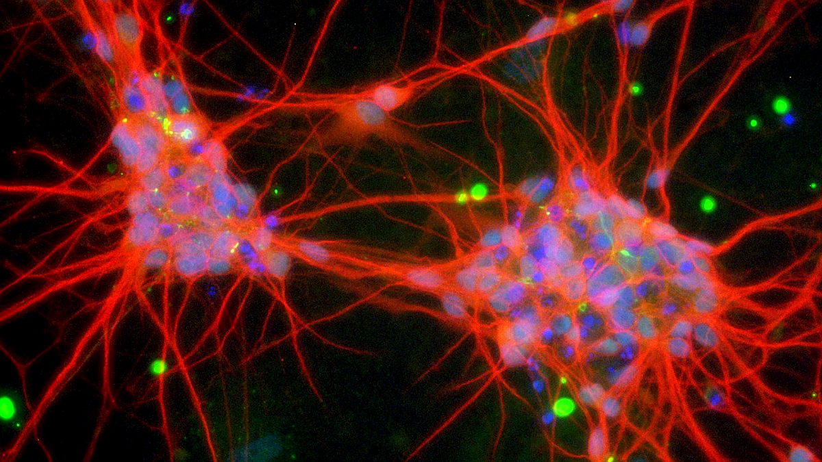
Confocal image of neurons differenciated from human embyonic stemm cells stained with Map2 (red), GAD67 (green) and Dapi
Electron Microscopy Unit (EMU)
Exemplary Image Material

Bachelor thesis of Jacqueline Heinen-Weiler in co-operation with AG Prof. Gunzer, Institute for Experimental Immunology & Imaging, University Duisburg-Essen Zeiss Crossbeam 540
This colored scanning electron microscopic image shows a microbial organism acquired during a collection of air particles.
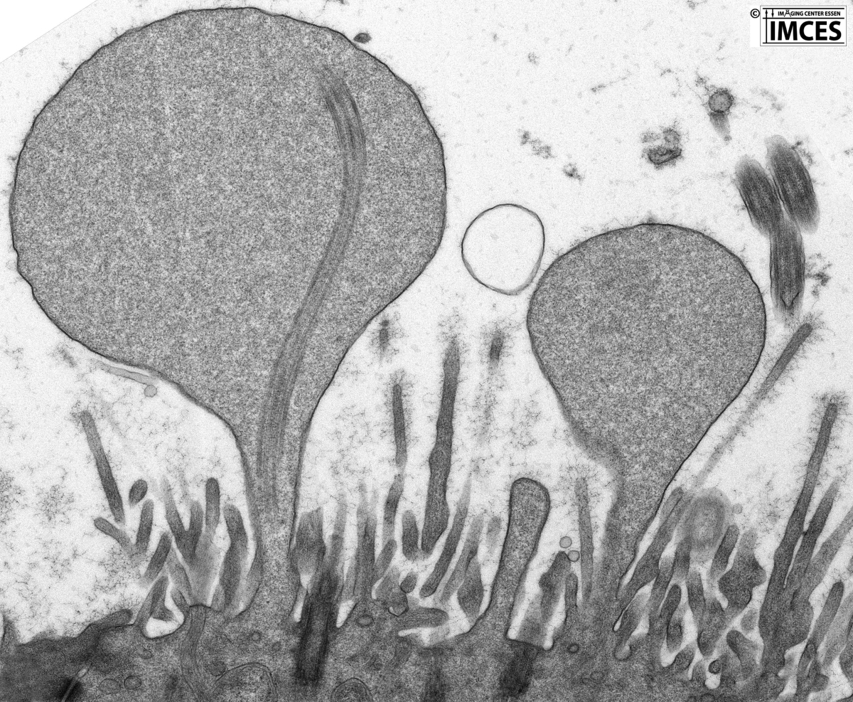
Co-operation with Prof. D. Theegarten, Institute for Pathology, University Clinic Essen JEOL 1400 Plus TEM
This transmission electron microscopic image shows pathologically blown-up kinocilia of a specimen of respiratory epithelium deriving from a biopsy of a patient after standard embedding and contrastation.
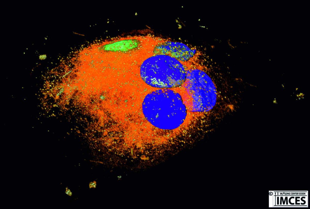
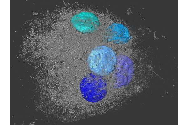
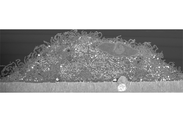
Master thesis of Frauke Uppenkamp at the EMU in co-operation with the Clinic for Orthopedics of the University Clinic Essen Zeiss Crossbeam 540
We see a 3D reconstruction of a multinuclear human osteoclast resulting from a nanotomography using FIB/SEM.
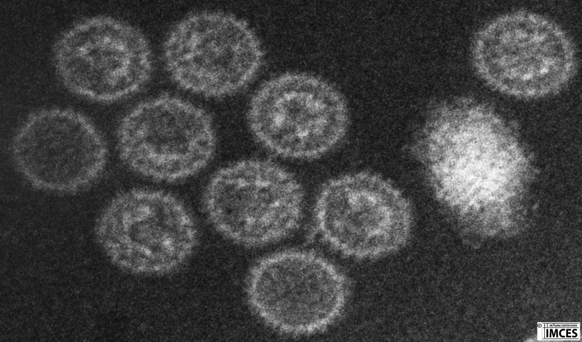
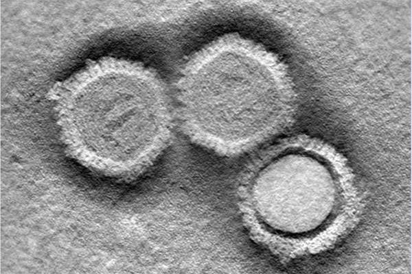
Master thesis of Sarah Borkowsky in co-operation with Prof. Mirko Trilling, Institute for Virology, University Clinc Essen JEOL 1400 Plus TEM
This transmission electron microscopic image shows capsids of murine cytomegaly virus (MCMV) after negative staining. Note that the virus particles are partially filled with DNA or empty.
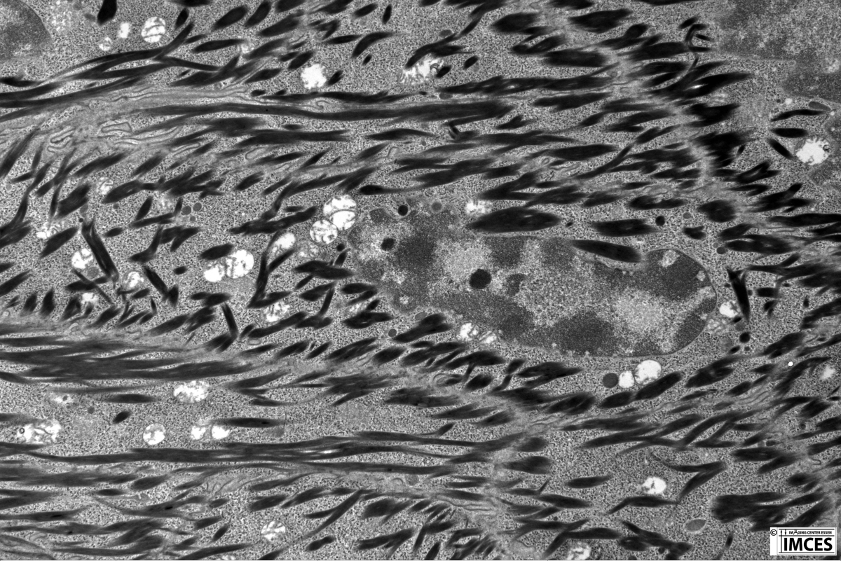
Co-operation with Prof. K. Willecke, University Bonn JEOL 1400 Plus TEM
This transmission electron image demonstrates keratin filaments in an epithelial cell of the mouse toe after standard embedding and contrastation.
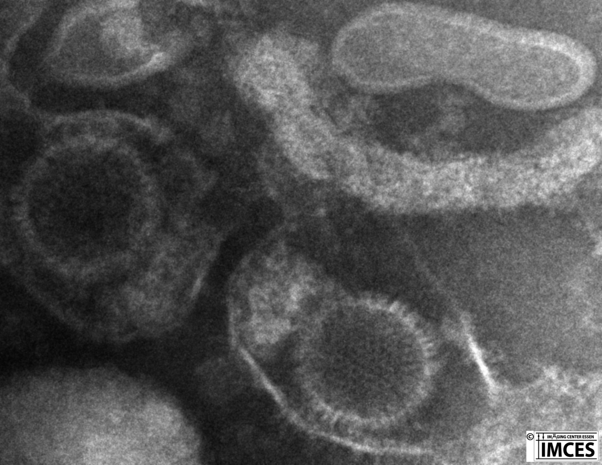
Master thesis of Sarah Borkowsky in co-operation with Prof. Mirko Trilling, Institute for Virology, University Clinc Essen JEOL 1400 Plus TEM
This transmission electron microscopic image shows capsids of murine cytomegaly virus (MCMV) after negative staining. Note the well preserved envelopes around the virus capsids.

Co-operation with PD K. D. Müller, Institute of Medical Microbiology, University Clinic Essen JEOL 1400 Plus TEM
This transmission electron micrograph demonstrates bacteria after standard embedding and contrastation.
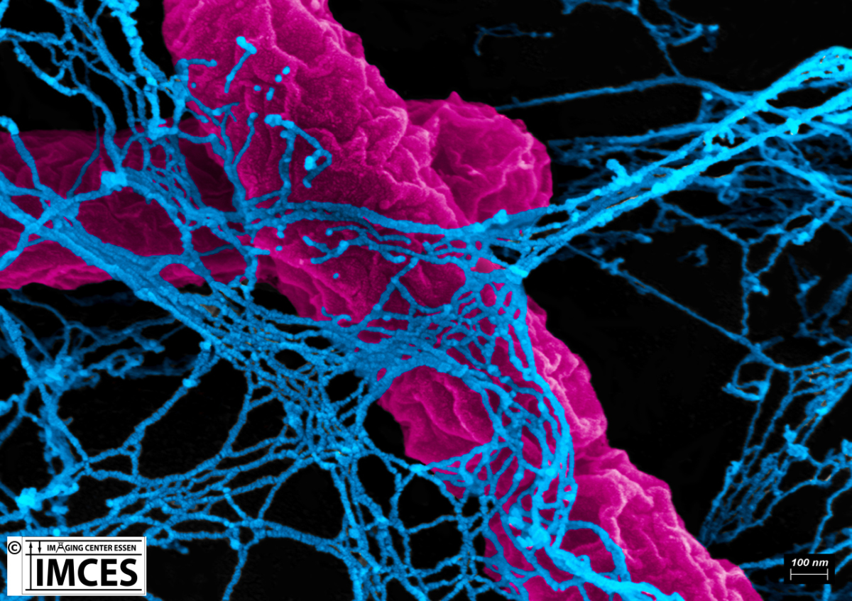
Bachelor thesis of Jacqueline Heinen-Weiler in co-operation with AG Prof. Gunzer, Institute for Experimental Immunology & Imaging, University Duisburg-Essen Zeiss Crossbeam 540
This colored scanning electron microscopic image shows Neutrophil Extracellular Traps (NETs) ejected from human neutrophilic granulocytes in which two E. coli bacteria are trapped.
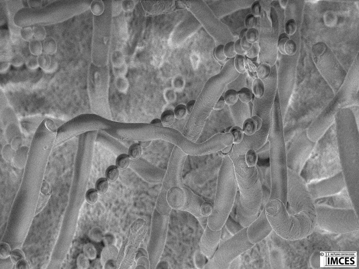
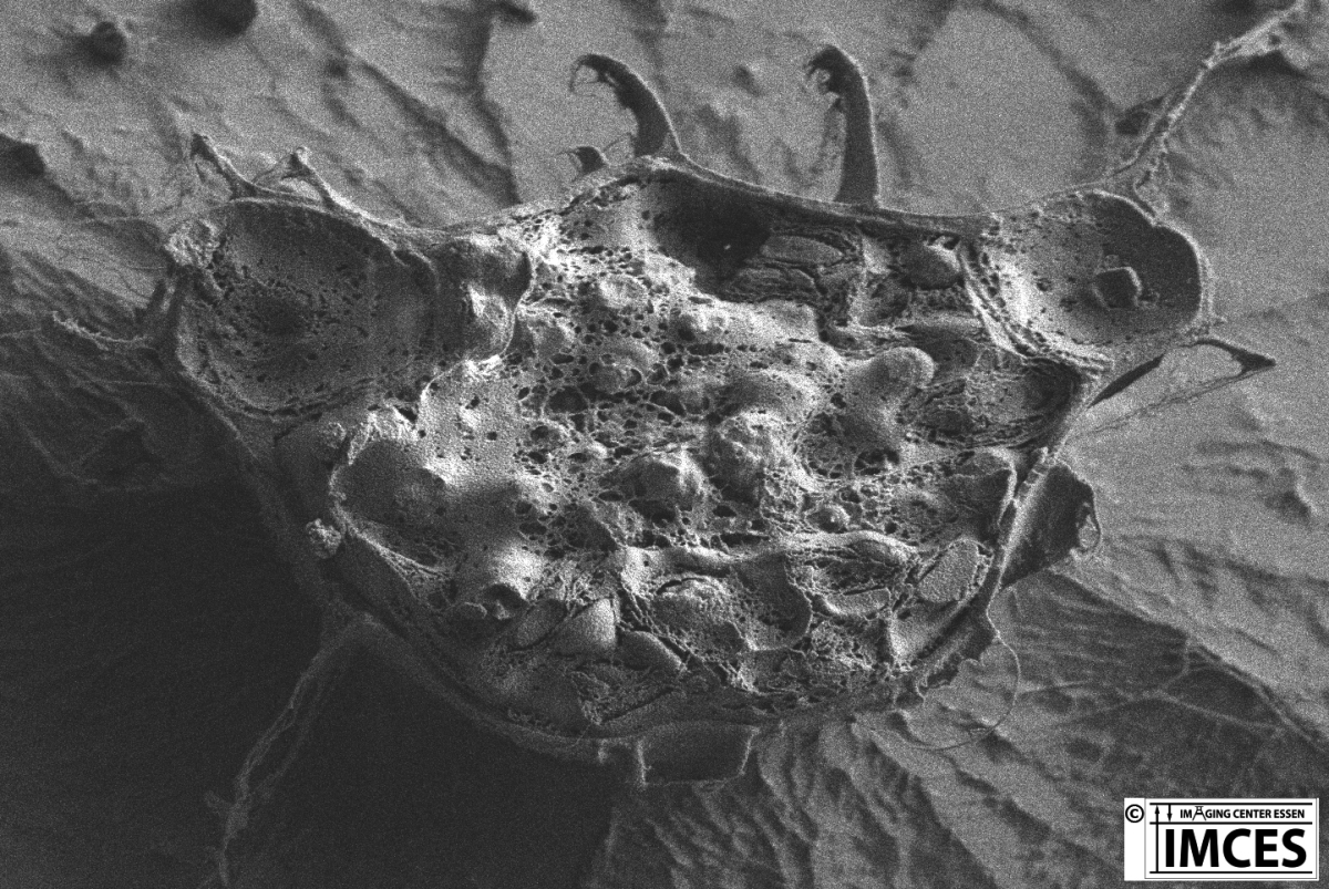
Bachelor thesis of Jacqueline Heinen-Weiler in co-operation with AG Prof. Gunzer, Institute for Experimental Immunology & Imaging, University Duisburg-Essen Zeiss Crossbeam 540
This image shows conidia and the dense mycelial network of Aspergillus fumigatus. The image was acquired using cryo scanning electron microscopy.
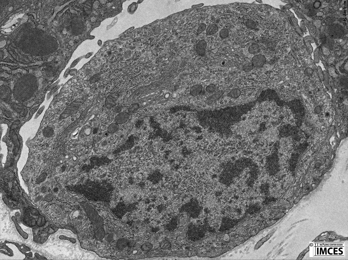
Transmission electron microscopic demo image JEOL 1400 Plus TEM
This transmission electron microccopic image demonstrates cell organelles of a calcitonin-producing cell of the thyroid gland.
Co-operation withSabina Marks (born Wodniok), CCAC Algae collection, Faculty of Biology, University Duisburg-Essen, Germany Zeiss Crossbeam 540
This cryo scanning electron microscopic image shows the cytoplasm and organelles of a "cracked" alga.
further images in preparation!
Exemplary Video Material
Master thesis of Frauke Uppenkamp at the EMU in co-operation with the Clinic for Orthopedics of the University Clinic Essen Zeiss Crossbeam 540
This motion picture shows a reconstructed osteoclast with colored nuclei in different views.
Master thesis of Frauke Uppenkamp at the EMU in co-operation with the Clinic for Orthopedics of the University Clinic Essen Zeiss Crossbeam 540
This animation demonstrates a series of sections through an osteoclast generated using the focused ion beam method (FIB).
Bachelor thesis of Kai Liebig at the EMU in co-operation with PD Dr. A. Krawczyk, Insitute for Virology University Clinic Essen JEOL 1400 Plus TEM
This is an animation through 3 tomographically reconstructed Herpes simplex viruses
further demonstrations of animations in preparation!