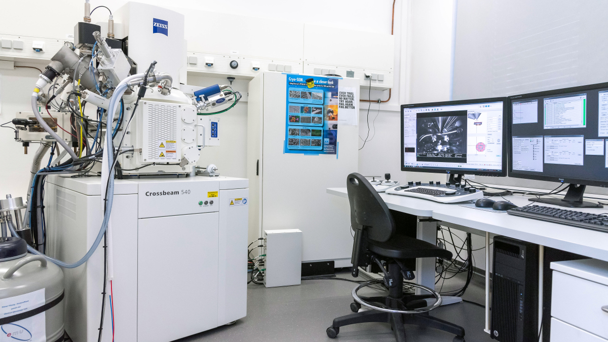
ZEISS - Crossbeam 540
Room 1039 1st floor MFZ building
Our high-end cryo-SEM with a Gemini column and a Schottky field emitter is equipped with a Ga+++ focused-ion beam system and platinum as well as carbon gas injection systems for quality enhancement of images. There are in-lens-, chamber- and energy selective detectors and a 6 axis stage for specimens which can be observed by 2 in chamber CCD cameras.
Besides conventional scanning electron microscopy for visualization of surfaces of specimens with a maximal resolution of 0.7 nm at 30KV, also transmission electron microscopic specimens may be investigated by means of the STEM detector. By means of our cryo-SEM preparation system it is possible to investigate shock-frozen samples, e.g. fresh biopsies without any fixation and sputtering artefacts in cryo-scanning electron microscopy.
The most important feature of the SEM, however, is the possibility to perform FIB-SEM investigations. A specimen is partly vaporized by means of a gallium ion beam after in chamber sputtering in a way that a hole with a plain wall is deeply cut into the sample. The flat surface of the wall then is digitized by SEM scanning. Then a next lamella of the surface is vaporized with the ion beam resulting in a next exactly parallel "section" which is scanned again. By repeating this procedure several hundred or thousand times a huge stack of exactly parallel oriented serial sections can be generated with a minimal lamella distance of 20 nm. The Capella column hereby allows a point resolution of 3 nm. Acquired data stacks then can be used for reconstruction of larger ultrastructures like complete mitochondria. The FIB is controlled by the image acquisition software Zeiss Atlas 3D which allows to generate 16-bits of gray images with resolution up to 40.000 x 50.000 pixels. Our workstation may be used for later generation of 3D-reconstructions from segmented structures and their high-end quality visualization.
The SEM is further equipped with the ZEN SEM 2012 software for correlative light and electron microscopy (CLEM). Hereby samples are first investigated by fluorescence microscopy and then scrutinized by SEM or STEM whereby the samples are adjusted in a way that the images acquired with both methods are exactly from the same region allowing over-projection for correlation of structures.
For long-time projects it is possible to work alone on this device after a sophisticated instruction.