Caliper Lumina
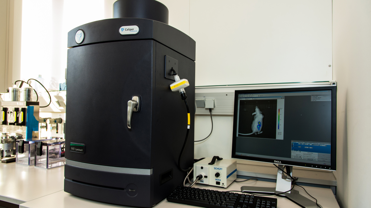
Light MicroscopySmall animal in vivo imaging
Download our operating protocol.
Technical Details:
System: Lumina II
Small animal, in vivo, quantitative, fluorescence and bioluminescence, molecular imaging system.
Software: Living Image
Excitation wavelengths: high resolution 35 nm bandwidth centered at: 430, 465, 500, 535, 570, 605, 640, 675, 710, 745.
Standard filter set and fluorophores:
|
Label |
Excitation bandpass (nm) |
Emission bandpass (nm) |
background bandpass (nm) |
Fluorophores |
|---|---|---|---|---|
|
Green |
445-490 |
515-575 |
410-440 |
GFP, EGFP, FITC |
|
Red |
500-550 |
575-650 |
460-490 |
DsRed2-1, 605 Qdot Bioconjugate |
|
Far Red |
615-665 |
695-770 |
580-610 |
Cy5.5, Alexa 660, Alexa 680, 705 QDot Bioconjugate |
|
NIR |
710-760 |
810-875 |
665-695 |
Indocyanine green (ICG), 805 Qdot Bioconjugate |
CCD Camera: Andor Technologies scientific grade, back-thinned, back-illuminated, large format CCD.
|
CCD Camera |
Specification |
|---|---|
|
Sensor type |
Back illuminated |
|
CCD Format |
1024 X 1024 |
|
Pixel Dimensions (microns) |
13 X 13 |
|
Quantum Efficiency |
>85% 400-700 |
|
Readout noise |
<= 2 e RMS |
|
Dark Charge |
<0.0015 e/pix/sec |
|
CCD Digitization |
16 bit |
|
Cooling |
Thermoelectric |
Capacity: Up to 5 mice or 2 Rats
Gas anesthesia system: The isoflurane anesthesia system allows for the anesthesia of up to five mice in the imaging chamber during imaging. There is a separate chamber for inducing anesthesia prior to imaging.
Accessories:
Emission filter options:
|
label |
Emission bandpass 20nm, centered at (nm): |
Fluorophores |
|---|---|---|
|
500 Series |
500,520,540,560, 580, 600 and 620. |
GFP, YFP and PKH26 |
|
700 Series |
720, 740, 760, 780, 800, 820 and 840. |
ICG and Xenofluor 750 |
Lens options: XFOV-24 lens attachment for expanded field of view (5 mice).