Microscopy
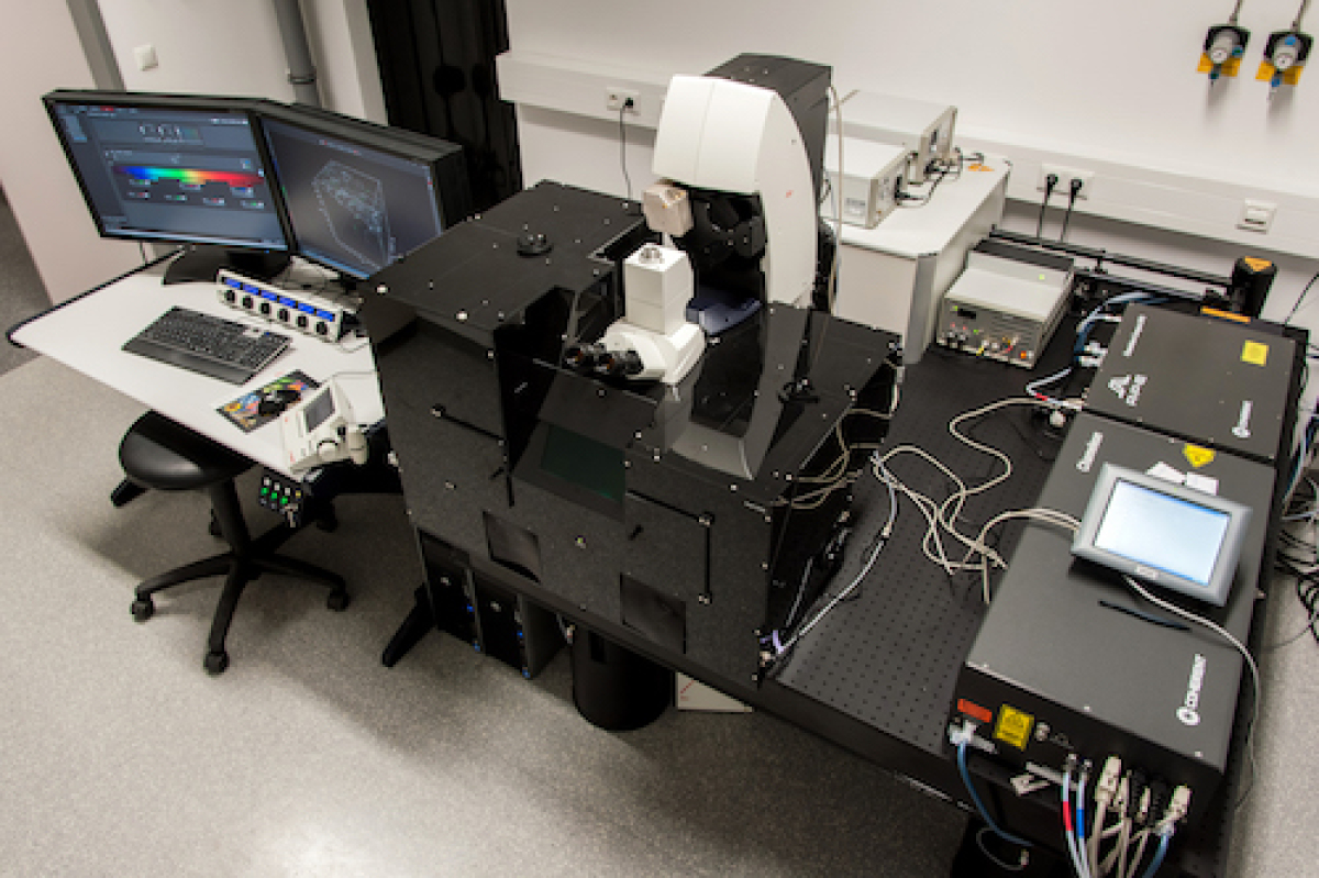
Light sheet fluorescence microscopy
Light sheet fluorescence microscopy (LSFM) is a novel method allowing the digital reconstruction of intact organs or whole animals with sub-cellular resolution within minutes. Via chemical procedures, samples have to be optically cleared to overcome opacity, which is generally limiting penetration depth in all types of fluorescence microscopy. The cleared samples are scanned with a LaVisionBioTec (Bielefeld, Germany) UltraMicroscope with an Olympus MVX10 zoom microscope body, an LVBT Laser module, an Andor Neo sCMOS camera with a pixel size of 6.5 x 6.5 µm2 and detection optics with an optical magnification range of 1.26 x - 12.6 x and an NA of 0.5. 3D rendering of LSFM data is performed via IMARIS software (Bitplane, Switzerland).
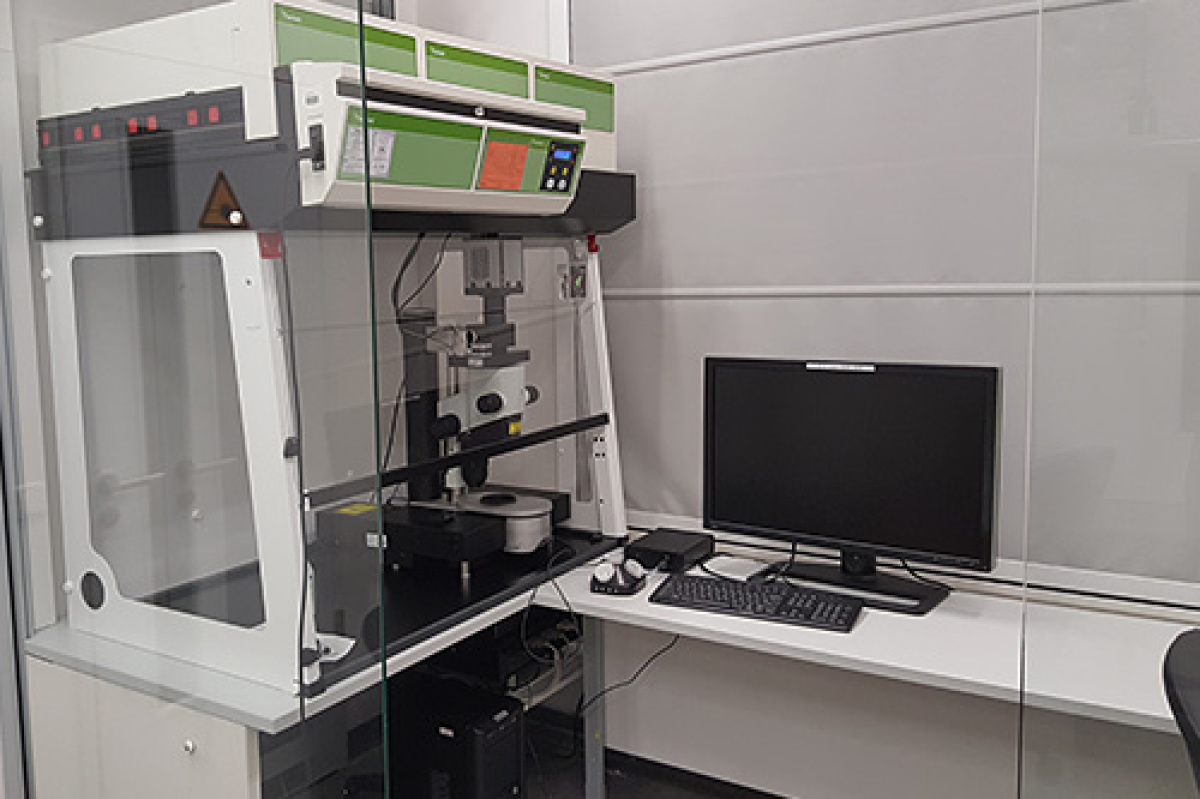
Confocal and multi-photon microscopy
The Leica TCS SP8 is mainly used and constructed for intravital microscopy or thick tissue slices. Due to its pulsed 2-photon laser, it allows the deep tissue penetration needed for these experiments. It is equipped with Acousto-Optic Tunable Filter (AOTF) and an Acousto Optical Beam Splitter (AOBS), gated Hybrid Detector (HyD) and is also able to measure Fluorescence Lifetime Imaging (FLIM). Yet, we also perform confocal imaging of cleared tissue or histological samples on a variety of tissues.
The microscope is upright and includes an x-y motorized stage. For the z direction, the objective is movable over a distance of 13 mm. For intravital microscopy, the complete setting can be provided, including isofluran-oxygen intubation narcosis, a monitoring system to control body temperature, blood pressure etc. and several stages to fix and heat the animals. Thereby, we can analyze dynamic immune cell interactions in several immunological approaches and disease models. We are experienced in the visualization of a variety of tissues including long bone, skull and lung as well as lymph nodes, brain and tumor imaging.
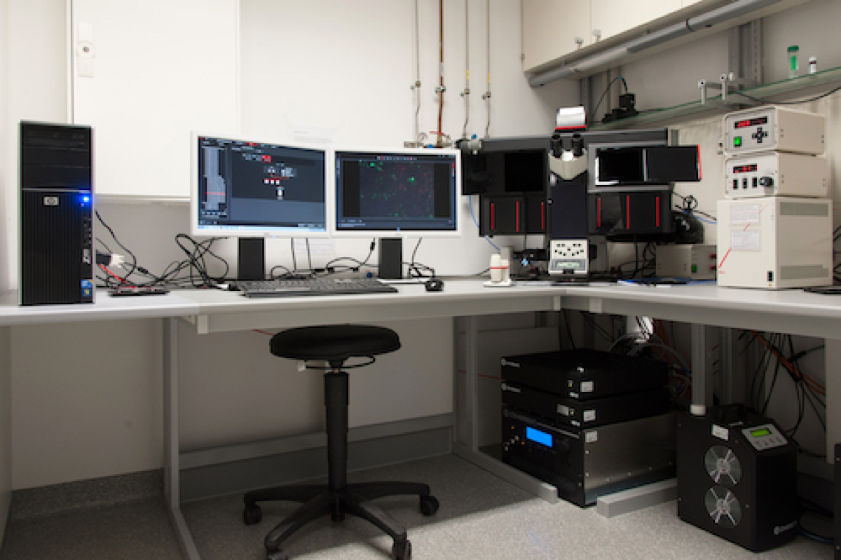
Time-lapse and fluorescence widefield microscopy
At our institute we are using our time-lapse fluorescence widefield microscope for various applications. Equipped with a fluorescence unit and an ultra-fast electric shutter we are able to perform not only widefield but also fluorescence microscopy including live cell imaging. Using this technology we are monitoring cell movement, fungal hyphae growth and phagocytosis of labeled particles. We are using a Leica DMI 6 equipped with two cameras for analysis - DFC365FX and DFC295. We use lenses ranging from 10x to 100x magnification and a fluorescence unit allowing us to analyze fluorescent dyes and proteins from violet to red.