See the voting results for the "Life Beyond Vision" Calendar 2026 here.
"Life beyond Vision" - Calendar 2025
Pictures of the IMCES calendar 2021
January 2021
February 2021
March 2021
April 2021
May 2021
June 2021
July 2021
August 2021
September 2021
October 2021
November 2021
December 2021
Pictures of the IMCES calendar 2020
Pictures of the IMCES calendar 2019
Pictures of the IMCES calendar 2018
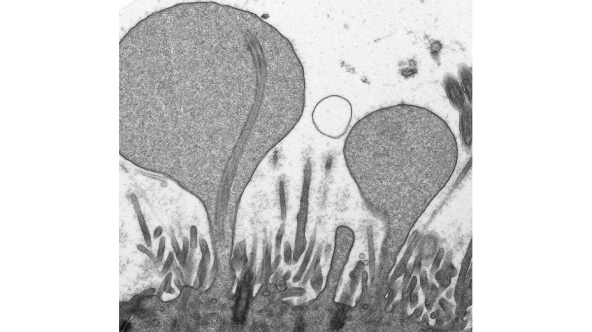
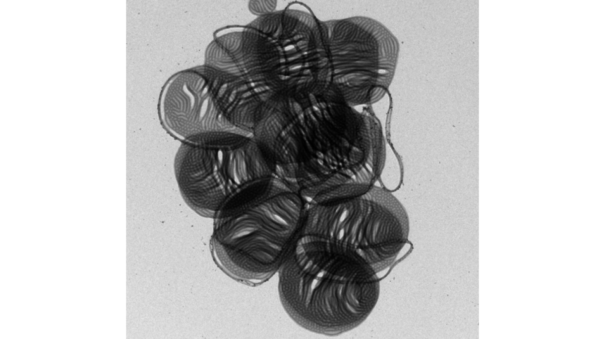
"Blown up” bleb-like protrusions of nose epithelial cells (Transmission electron microscopy)
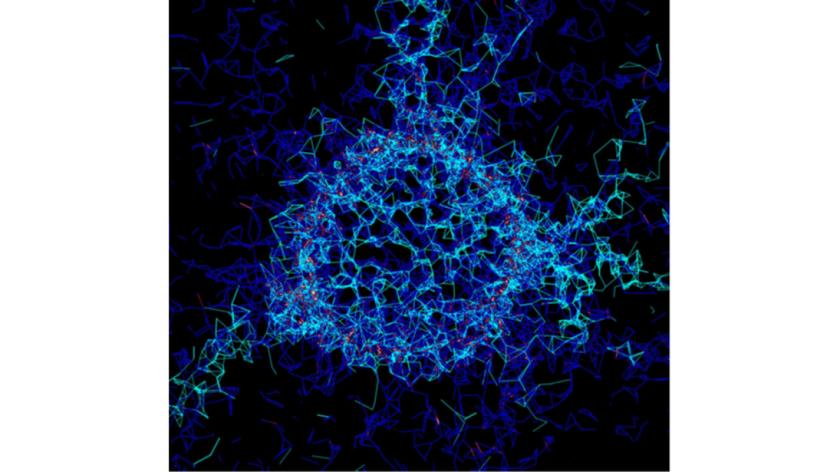
Graph-based mathematical reconstruction of a perineuronal net. Aggrecan mesh (cyan) and glycan network (blue) is shown. Interaction betweent the nets is depicted in red to white scale
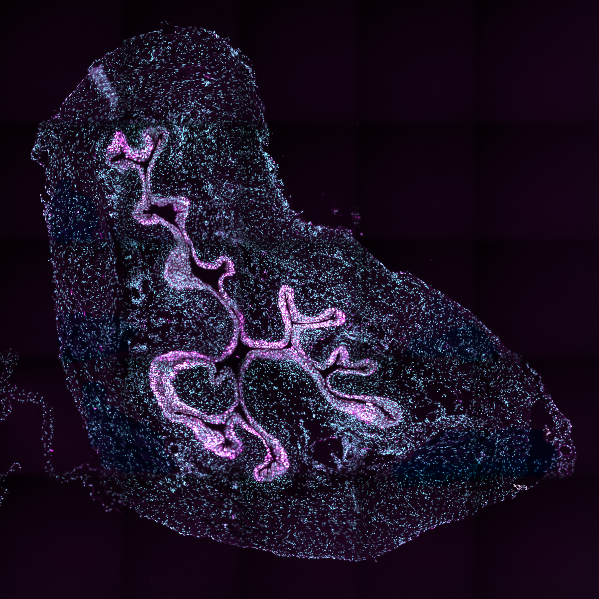
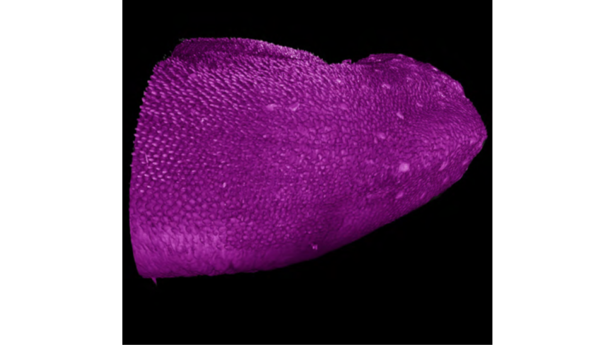
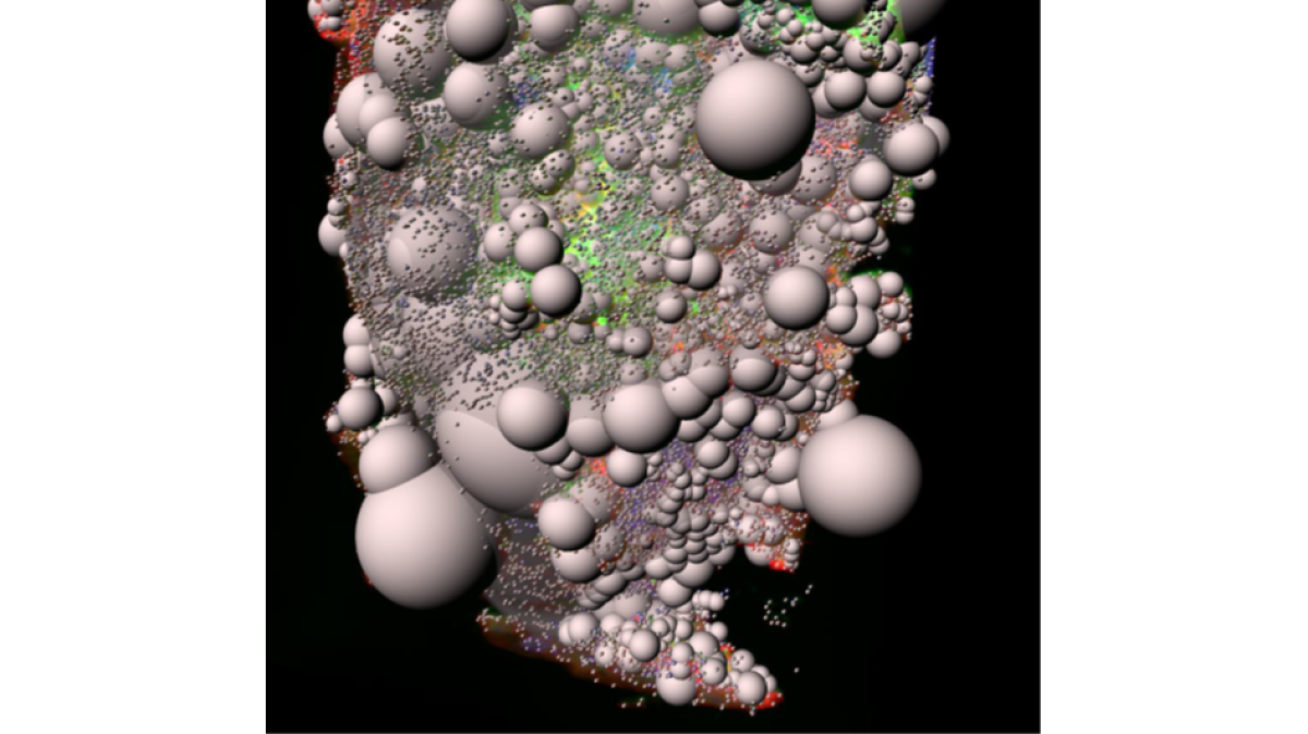
Light Sheet Fluorescence Microscopy (LSFM) – 3D surface rendering of a murine tongue tip
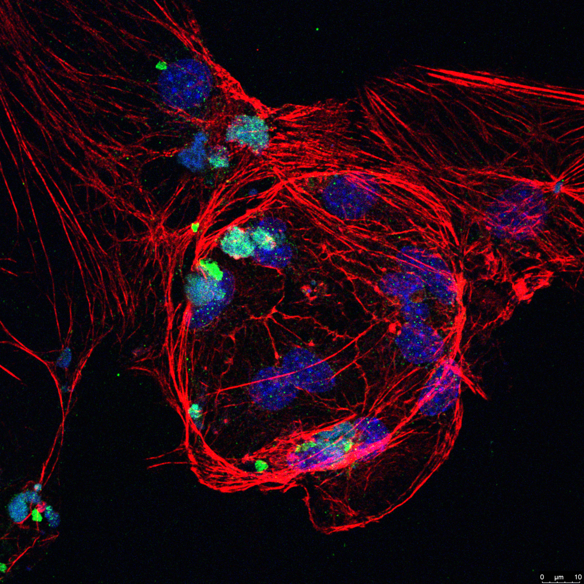
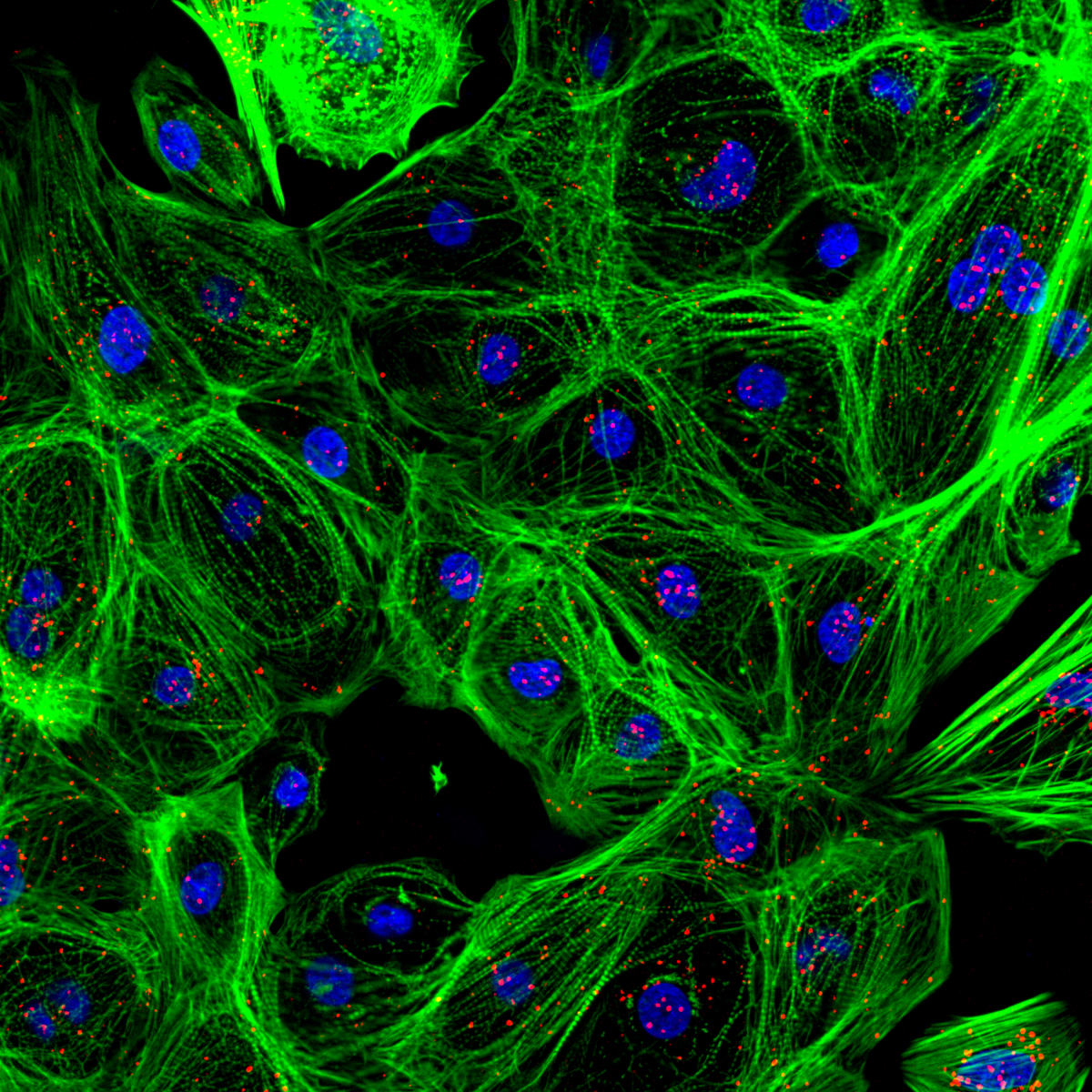
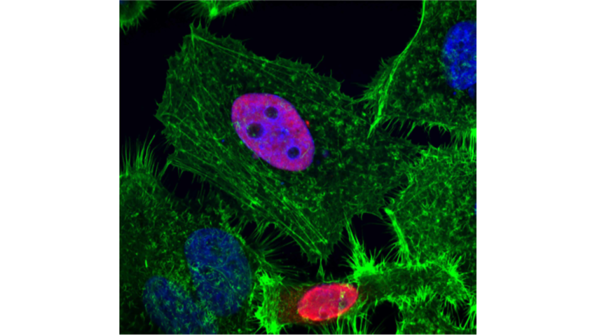
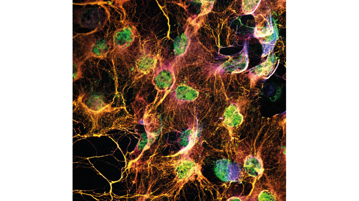
"Hela cells overexpressing the thyroid hormone receptor isoform TR Beta (red). Nuclei are labelled with DAPI (blue) and actin filaments are stained with Alexa Fluor 488 Phalloidin (green). (Confocal microscopy)
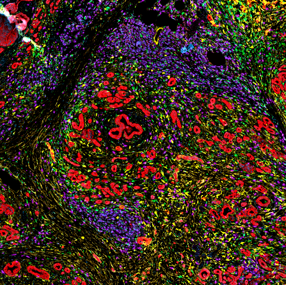
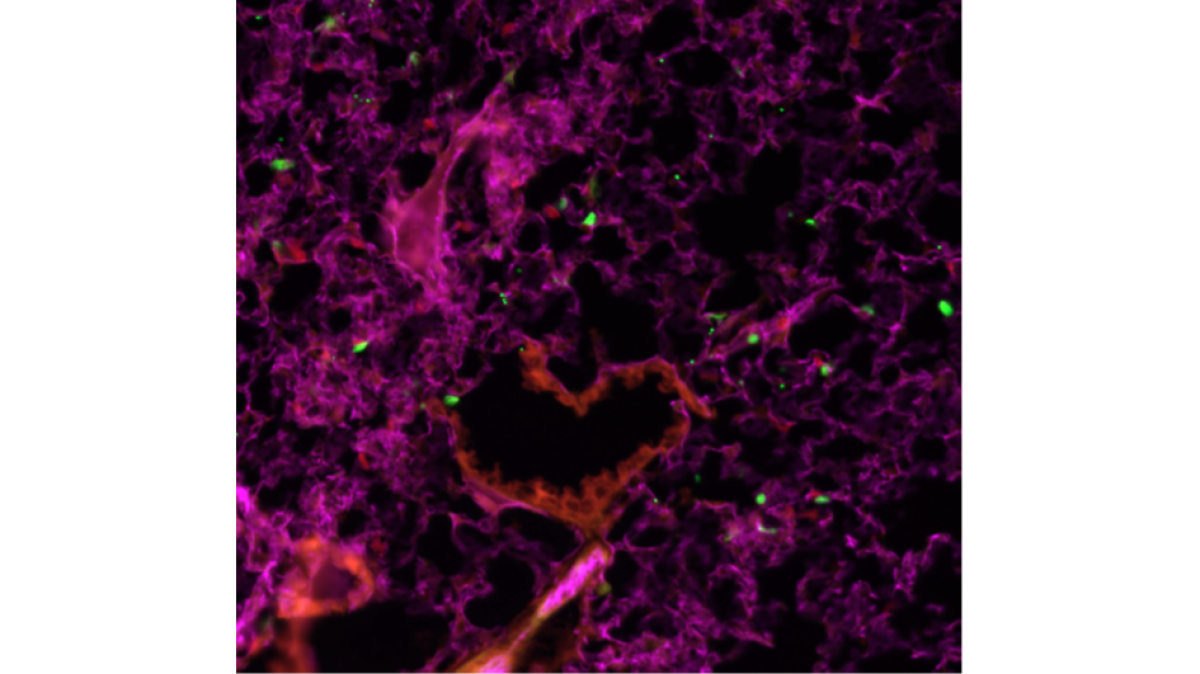
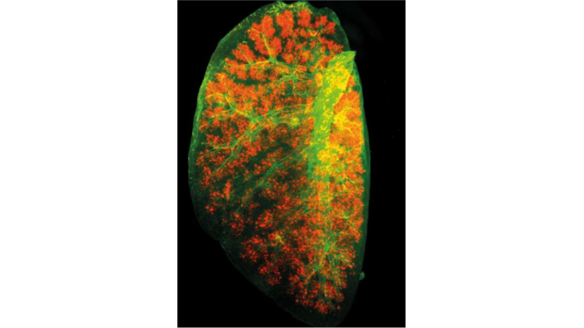
Epifluorescence microscopy – Immune cell recruitment into an infected murine lung Green – Macrophages and A. fumigatus conodia; Red – Neutrophils; Pink – Lung tissue
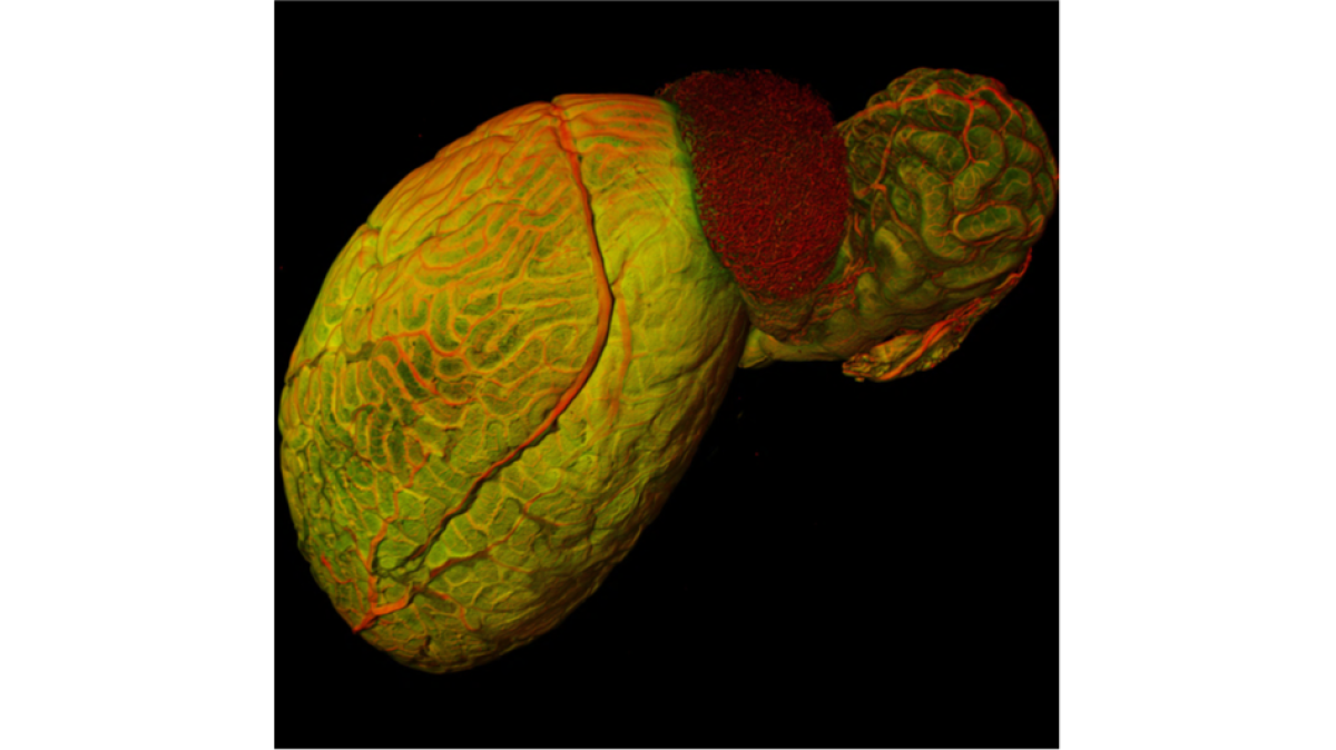
Light Sheet Fluorescence Microscopy (LSFM) – 3D visualization of a murine testicle and its vasculature
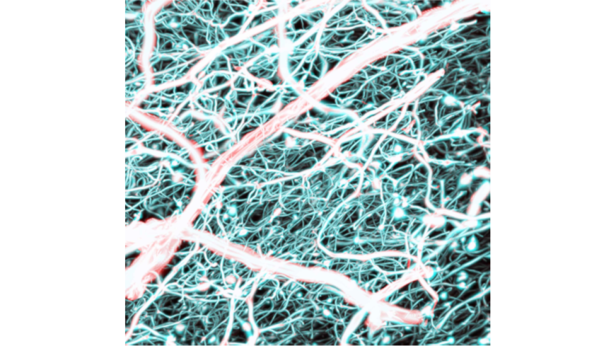
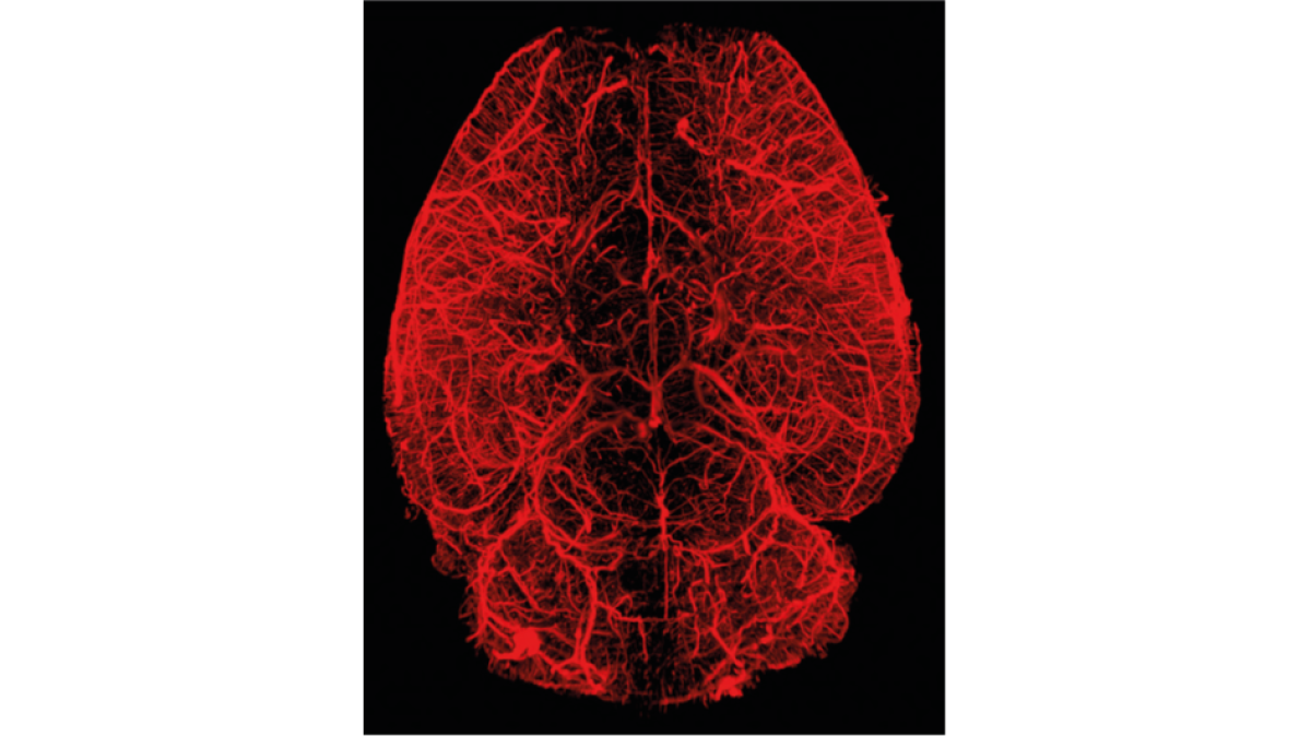
The cerebral vascular network of a mouse 56 days after experimental stroke. All blood vessels were labelled with FITC-conjugated albumin. (Light Sheet microscopy)
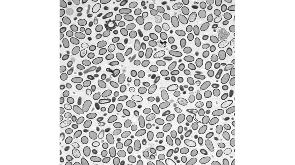
TEM image of triblock terpolymer nanostructures, prior cross-linked with OsO4 and dissolved in THF. The inner core appears black due to the high contrast of osmium and is surrounded by two coronal polymer layers below and above these grate-like structures
Light Sheet Fluorescence Microscopy (LSFM) – Vascular system of the murine brain
The cerebral vascular network of a mouse 56 days after experimental stroke. All blood vessels were labelled with FITC-conjugated albumin. (Light Sheet microscopy)
TEM image of triblock terpolymer nanostructures, prior cross-linked with OsO4 and dissolved in THF. The inner core appears black due to the high contrast of osmium and is surrounded by two coronal polymer layers below and above these grate-like structures
Light Sheet Fluorescence Microscopy (LSFM) – Vascular system of the murine brain
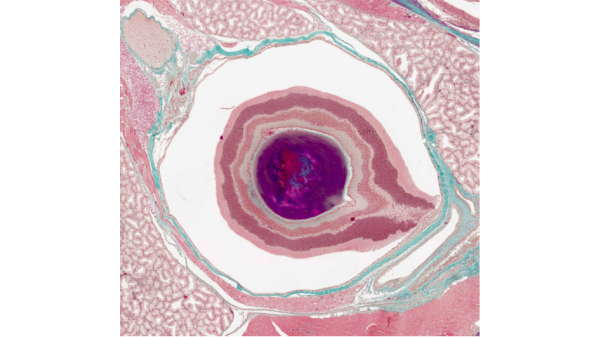
Coronal section of a mouse eye. Masson Trichrome (MTC) stained. (Light microscopy)
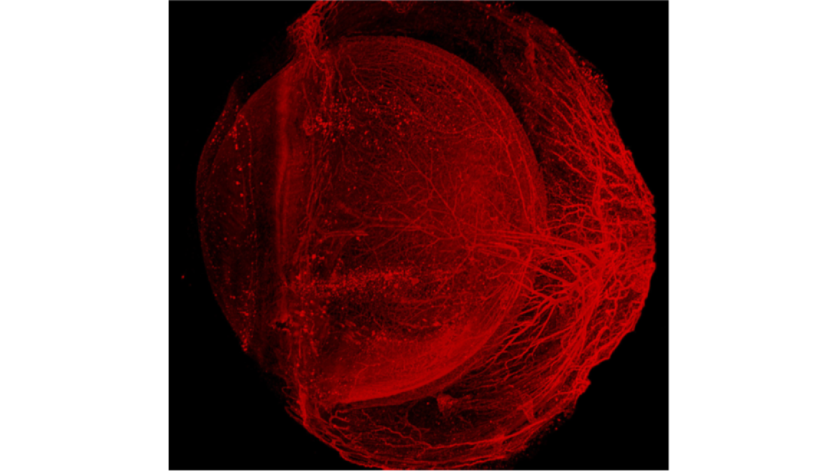
Entire vasculature of whole mouse eye (C57BL/6) immunostained (CD31, red) and cleared with EyeCi
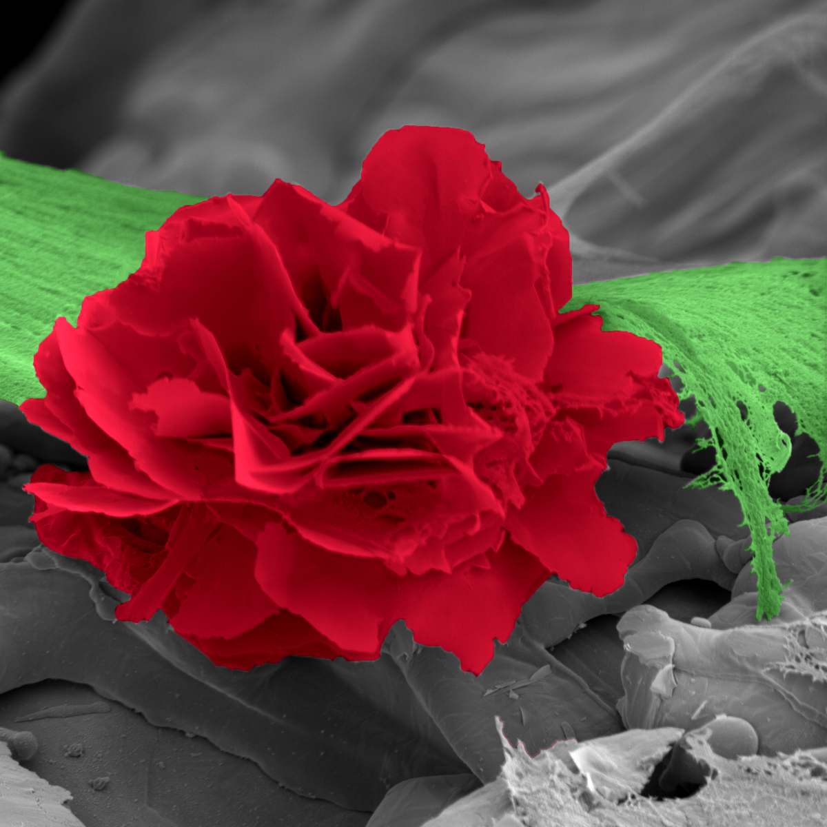
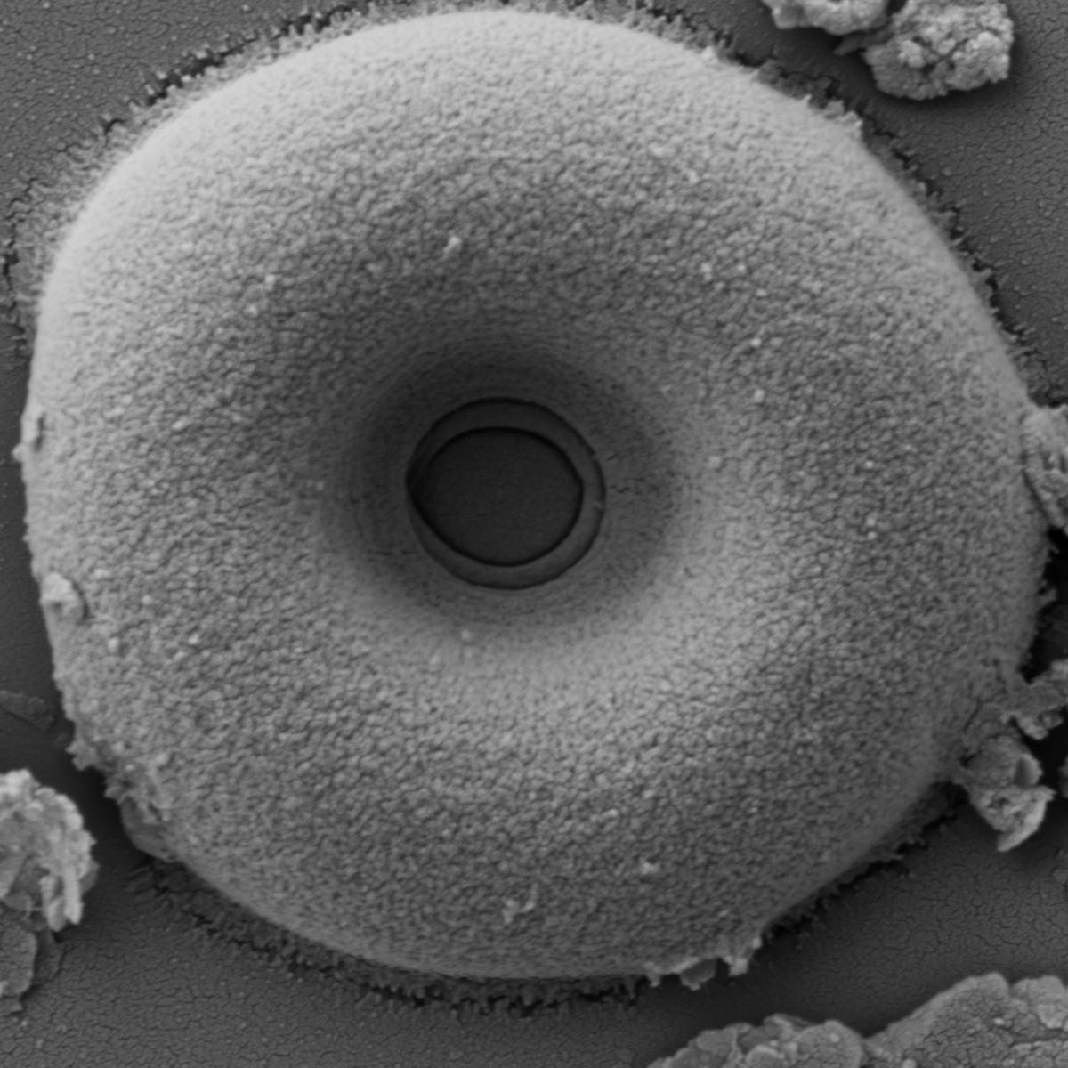
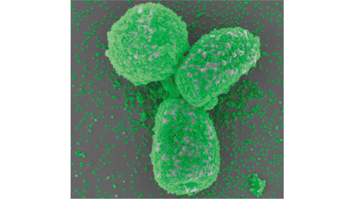
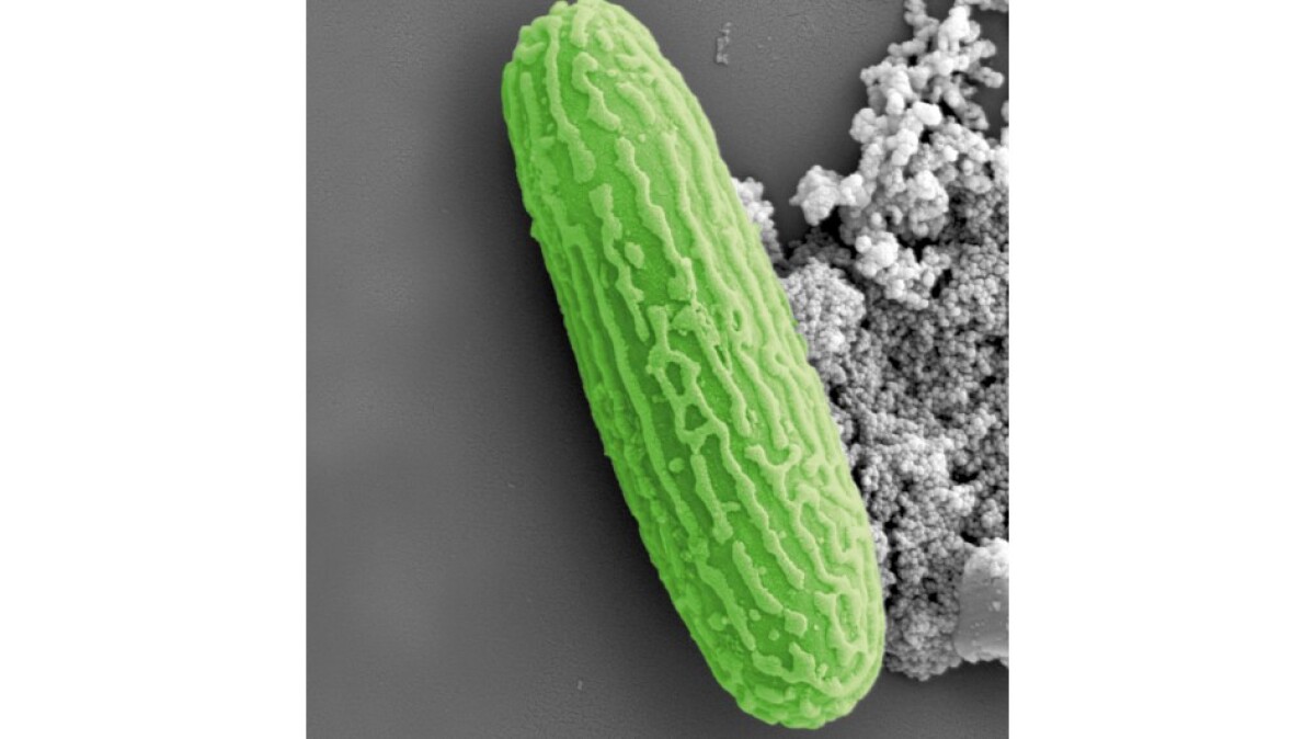
Scanning Electron Microscopy (SEM) – Spores of Aspergillus fumigatus entirely covered by silica nanoparticles
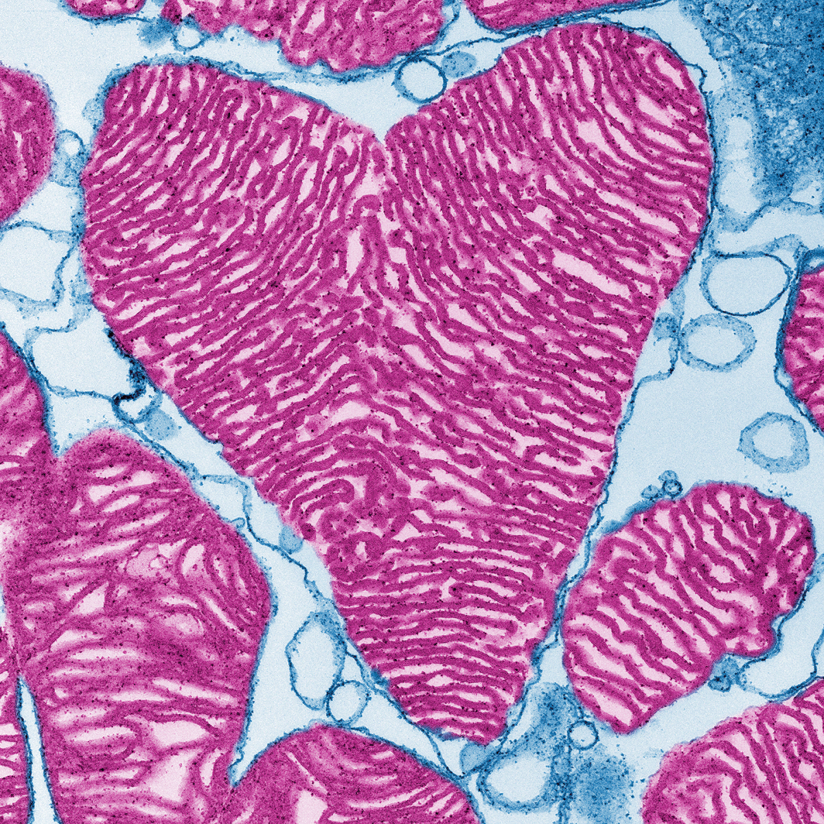
Mitochondria in a pig cardiomyocyte (Transmission electron microscopy)
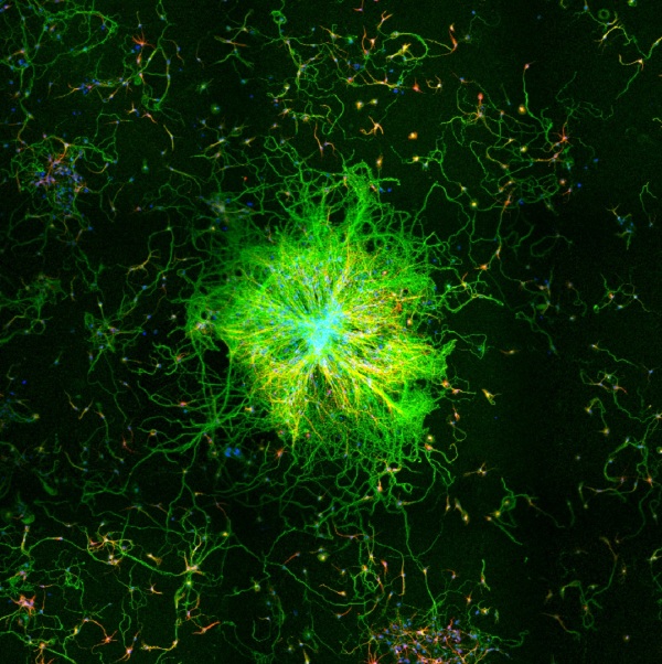
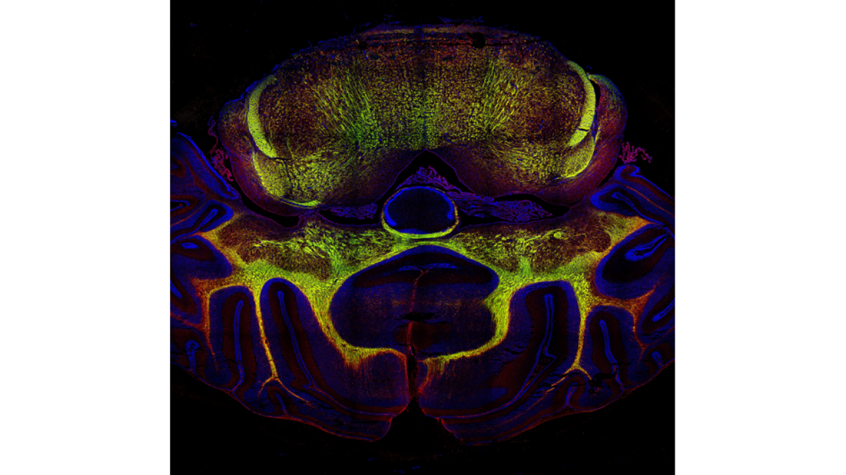
Laser Scanning Confocal Microscopy (LSM) – Expression of CEACAM1 (red) and MBP (green) in rhombencephalon and cerebellum of a 15 day-old rat
Sedia is exerovit, saperup tatius sapitia venimus. Sedia is exerovit, saperup tatius sa
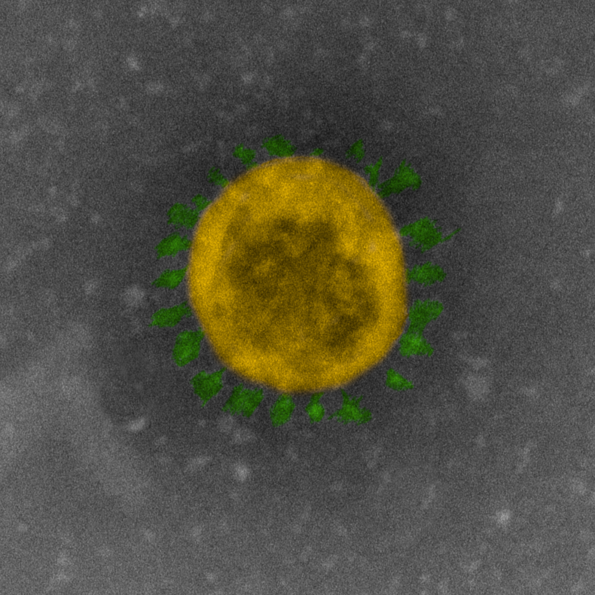
Topographic image of an airborne germ (Scanning Electron Microscopy)
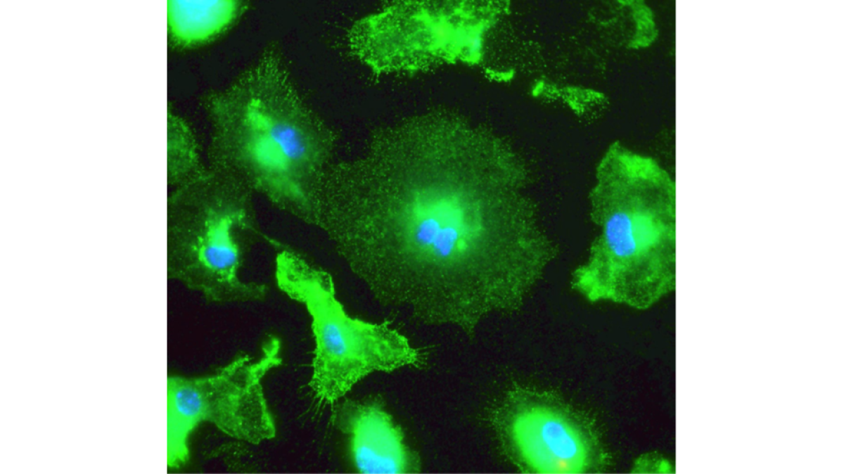
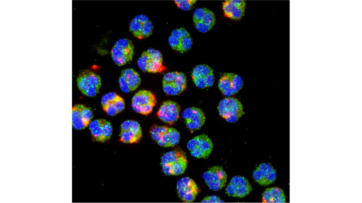
Primary mouse microglia stimulated in vitro with bacterial endotoxin / Iba-1 (green) and DNA (blue)
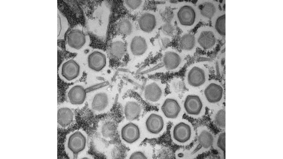
Transmission Electron Microscopy (TEM) – Enriched “giant virus” (Pithovirus lacustris) particles from an environmental soil sample
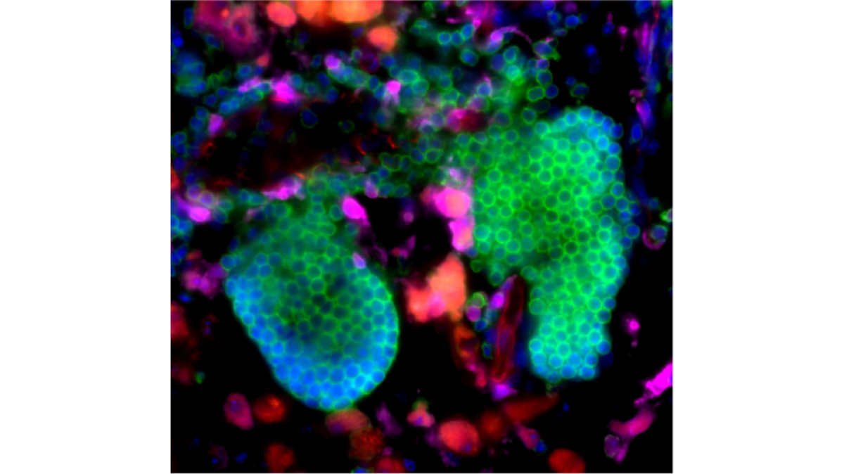
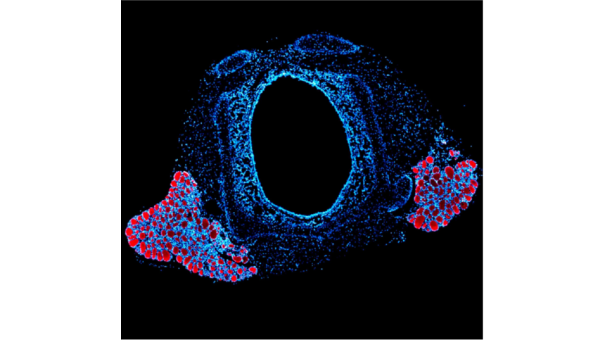
Cross-sectional image of the right and left lobe of a murine thyroid. Thyroglobulin is stained in red and nuclei are counterstained with DAPI (blue). (Fluorescence microscopy)
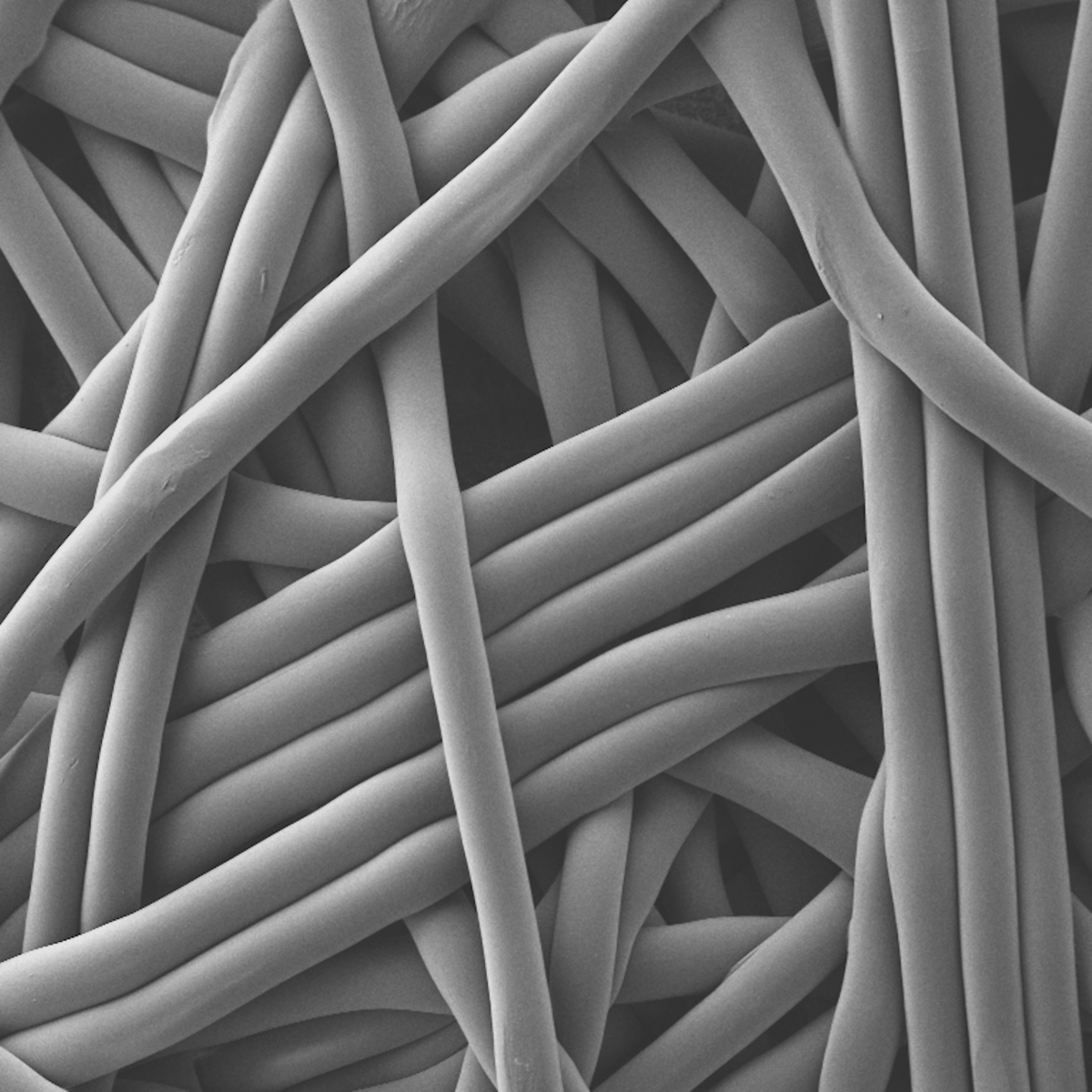
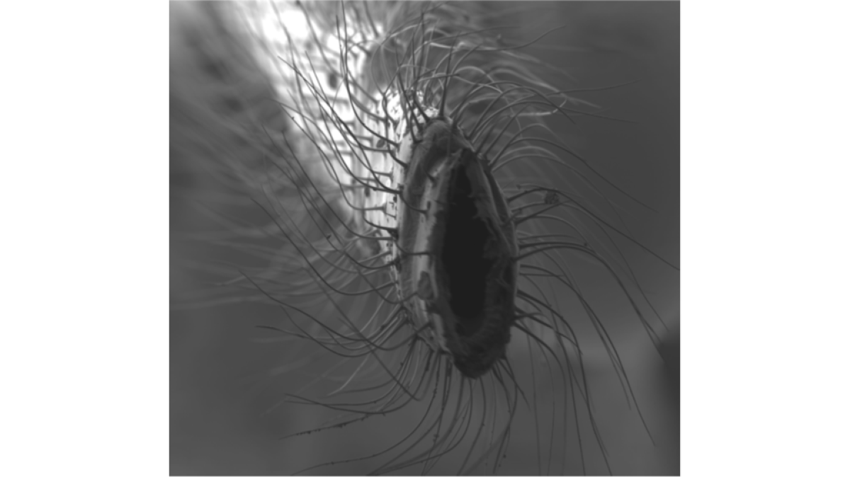
Scanning Electron Microscopy (SEM) – Topographic picture of a walking leaf’s (ophila melano) loose antenna
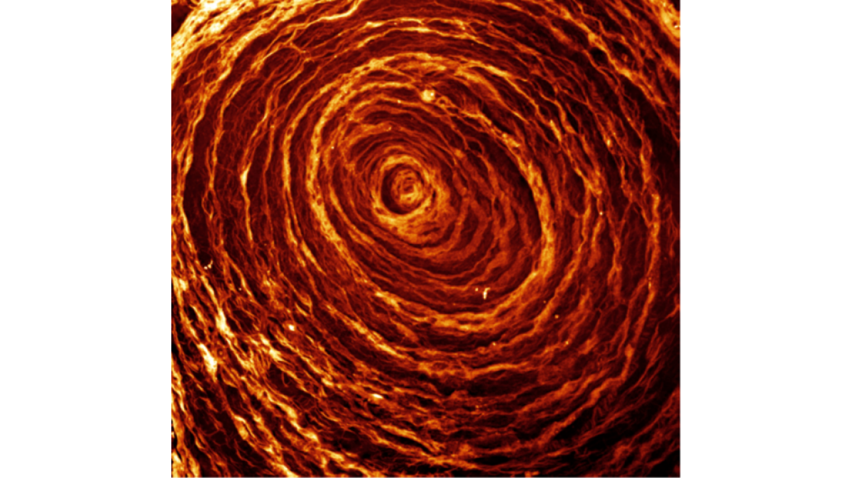
Structured Illumination Microscopy (SIM) – Fine Structure of Neutrophil Extracellular Traps
Neutrophil Granulocytes taking up gold nanoparticles ex vivo. (Confocal microscopy)
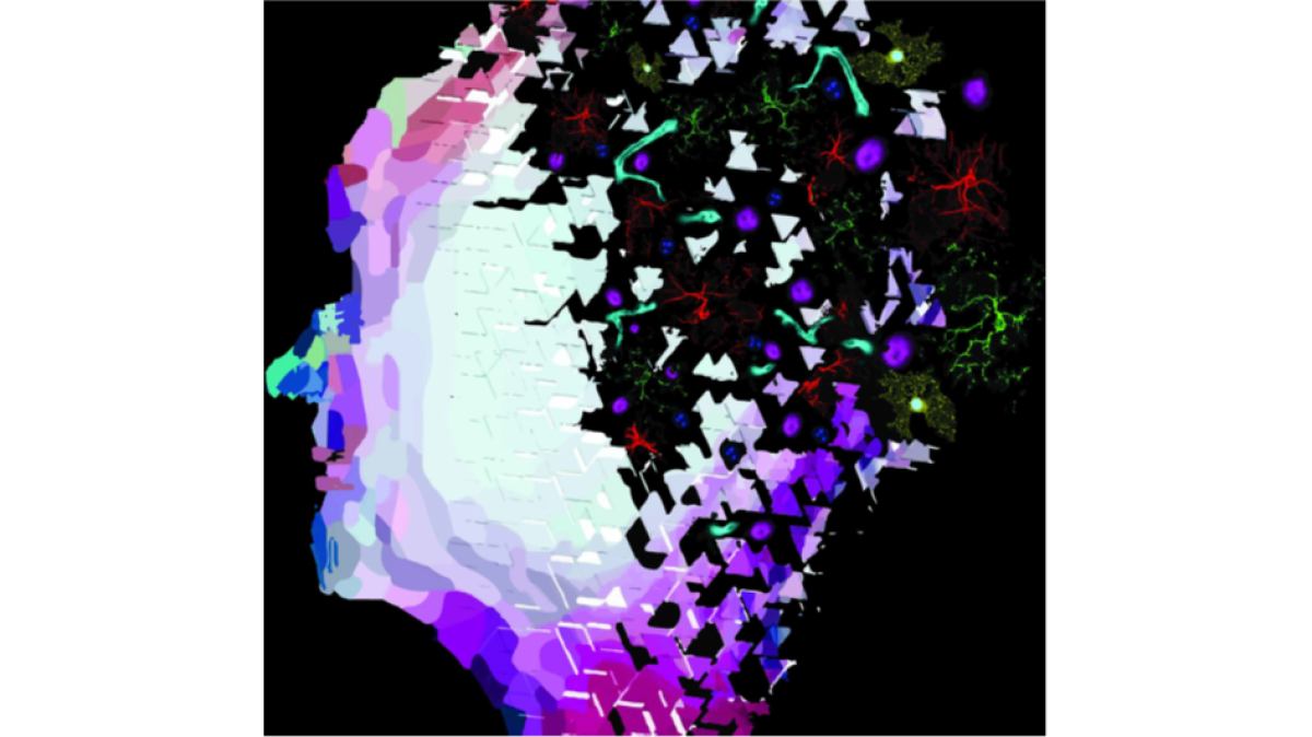
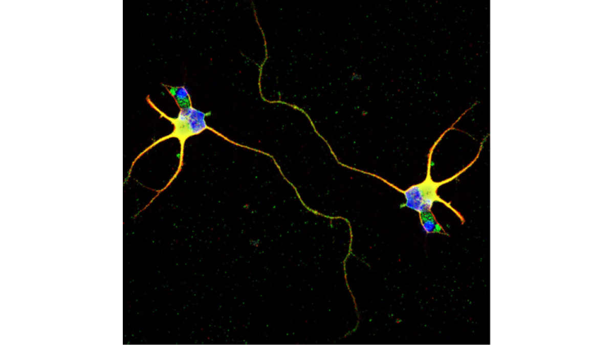
Single immature neuron under mirror transformation. Immunolabeling against Tau protein (red) and multivesicular bodies (green). Cell nuclei are labelled with DAPI (blue)
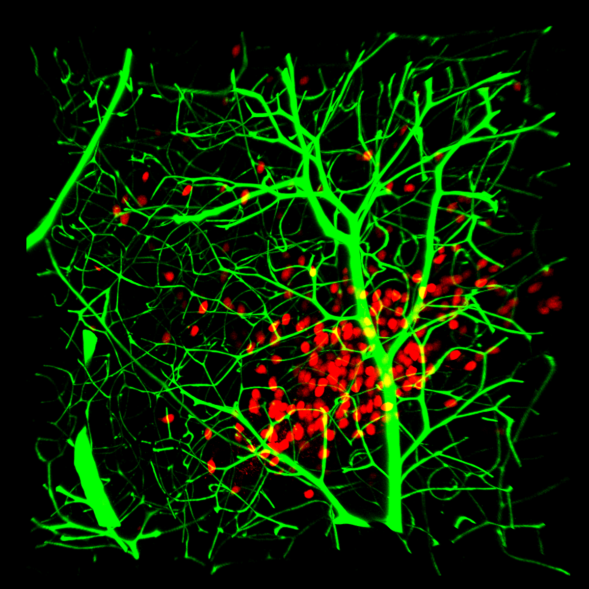
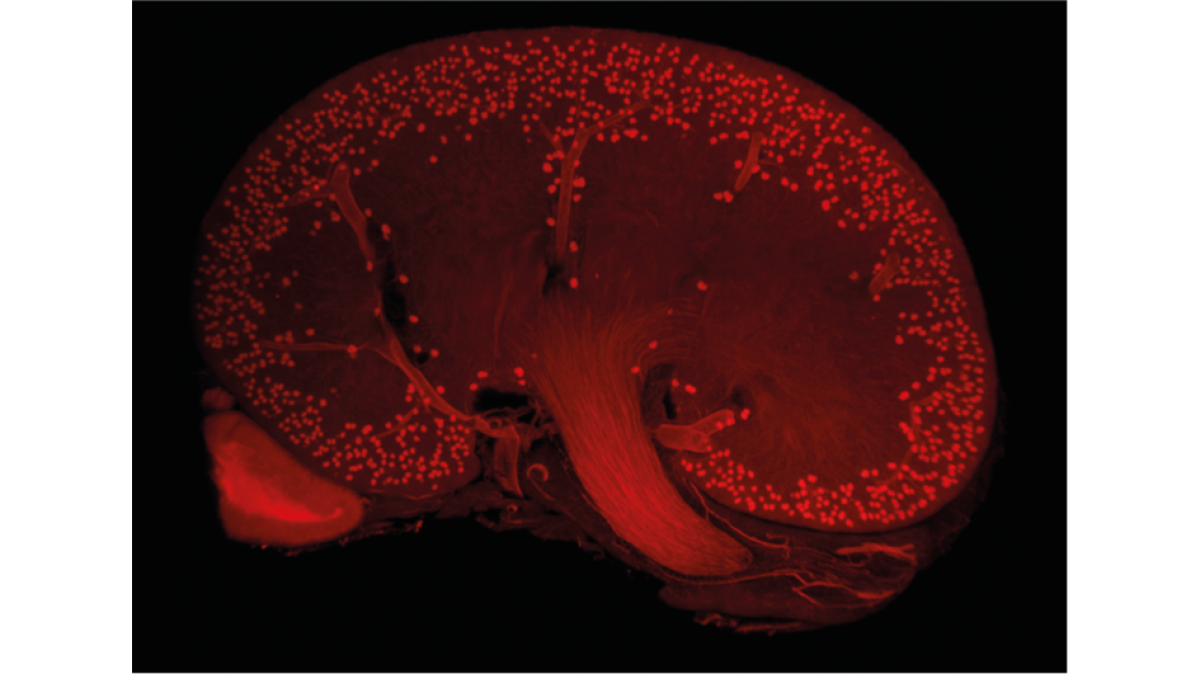
Light Sheet Fluorescence Microscopy (LSFM) - Midline optical section through a murine kidney with blood vessels stained in red
Vasculature of a murine heart following ischemia/reperfusion injury. (Light Sheet microscopy)
Virus particle formation inside an amoeba (Transmission electron microscopy)
Transmission Electron Microscopy (TEM) – A damaged cell body of the black yeast Exophiala dermatidis
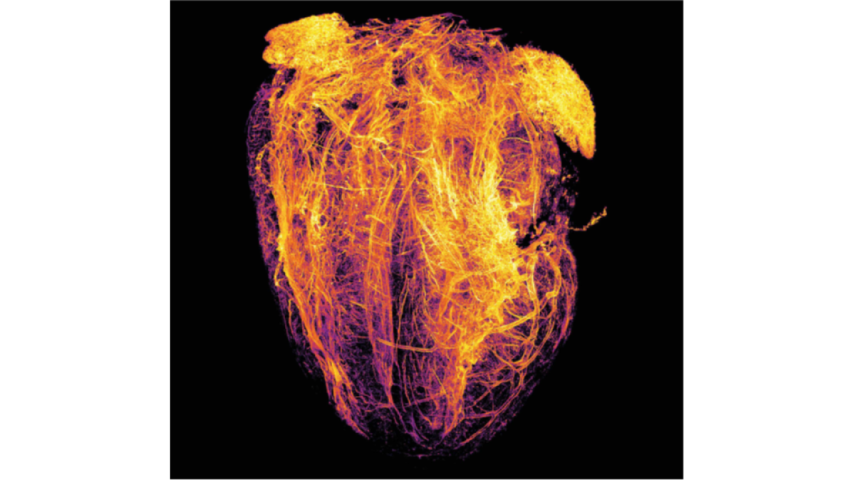
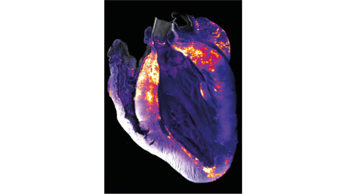
Light Sheet Fluorescence Microscopy (LSFM) – Heat-map visualization of a leukocyte infiltrate inside a murine heart
Mouse brain which suffered a stroke. Blood vessels are visible via CD31 (green) & platelets are shown via GP9 (red) antibody staining. The general tissue structure is shown in blue. (Light Sheet microscopy)
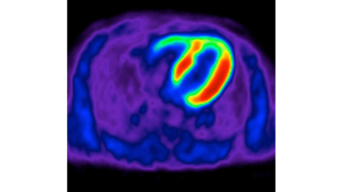
Transaxial view of a viability F-18 fluorodeoxyglucose (FDG) positron emission tomography (PET) study through the heart
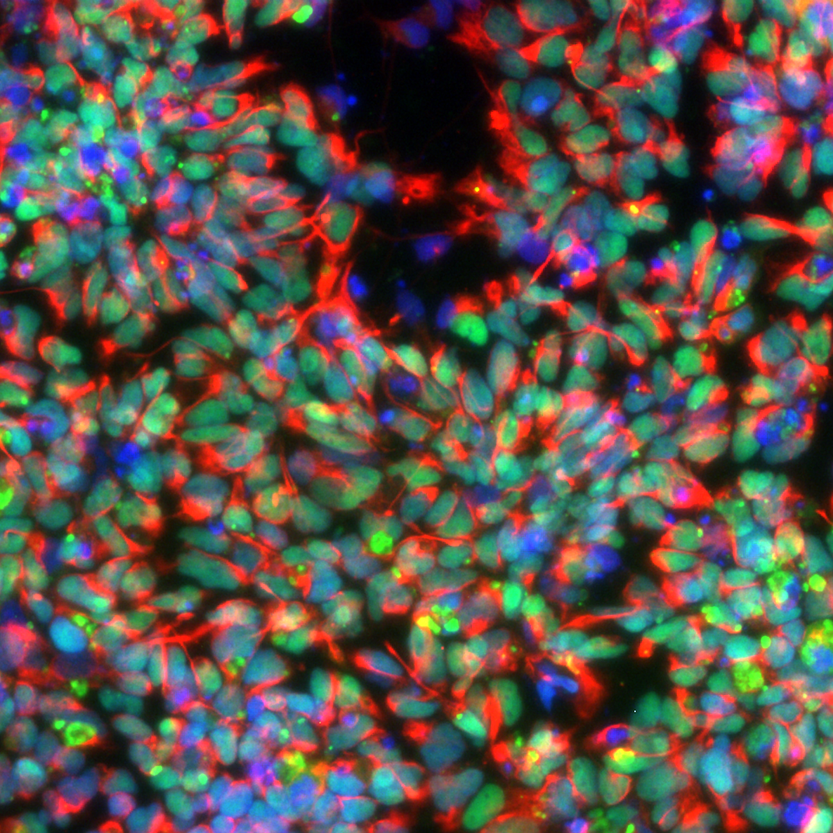
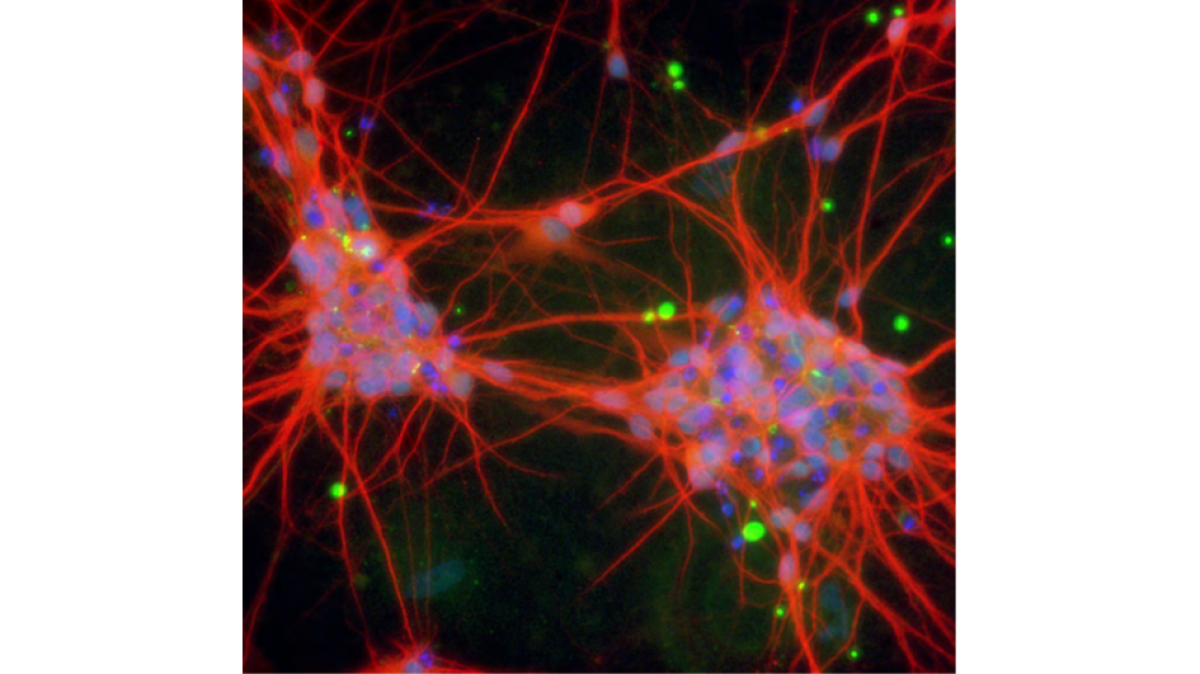
Laser Scanning Confocal Microscopy (LSM) - Neurons differentiated from human embryonic stem cells