Leica STED & FLIM
Light MicroscopyConfocal and superresolution microscopy
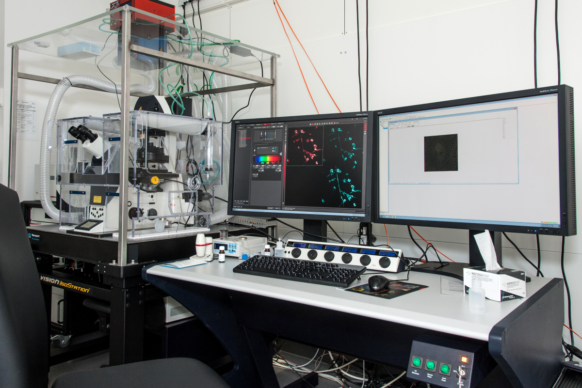
With the Leica SP8 and its choosable modules STED and FLIM you can nearly address any scientific challange which can be answeered by light microscopy. With our Leica SP8 you can perform normal light and fluorescence microscopy. The incubation chamber with a CO2 module easily allows live cell imaging using a resonant scanner equipped device. In case you want to investigate life time of fluorescence stimulated molecules you are welcome to use the FLIM module. In case you want to investigate your samples in more detail you can apply our 595 nm depletion laser in the STED module which under best conditions allows to gain a lateral resolution as tiny as 50 nms.
Technical Details:
Microscope:
Leica TCS SP8 fully automated epifluorescence confocal microscope, with AOTF and AOBS, white light laser (WLL2), gated HyD detection, gated STED,
and Fluorescence Lifetime Imaging - SMD SP FLIM.
Objectives:
10x (air) HCPL Fluotar with 11mm working distace and a numerical aperture of 0.3
20x (air) PL APO CS with 0.59mm working distance and a numerical aperture of 0.7
40x (oil) PL APO CS with 0.24mm working distance and a numerical aperture of 1.3
63x (water) HC PL APO CS2 with 0.3mm working distance and a numerical aperture of 1.3
63x (glycerin) HC PL APO motCORR CS2 with 0.3mm working distance and a numerical aperture of 1.3
100x (oil) HCX PL APO STED with 0.1mm working distance and a numerical aperture of 1.4
Confocal Scanner:
Field of View Scanner (FD 22mm, 8192X8192, 3600Hz) and Resonant (FD 13mm, 1024X1024, 12000Hz)
Confocal excitation lines:
Argon Ion (65 mW): 458nm,476nm, 488nm, 496nm, 514nm. Diode (50mW): 405nm.
White light laser WLL2 (1.5mW per line - 8 simultaneous possible): tunable from 470nm-670nm. picosecond pulsed, 78 MHz.
STED depletion laser (1.5W): 592nm.
5 Spectral Confocal Detectors: PMT and HyD (400nm-800nm):
1 internal SMD FLIM PMT, 2 internal PMTs, 2 internal HyDs (gated).
1 Confocal Transmitted Light Channel:
PMT
Super-resolution STED:
gated STED lateral FWHM 50nm, cw STED FWHM 80 nm, lateral. Normal confocal axial resolution.
STED Sample Preparation:
Protocol 1.
Fluorescence Lifetime Imaging Microscopy SP FLIM:
TCSPC module PicoHarp 300, Analysis software "SymphoTime" SPT2.