Transmission Electron Microscope JEOL 1400 Plus
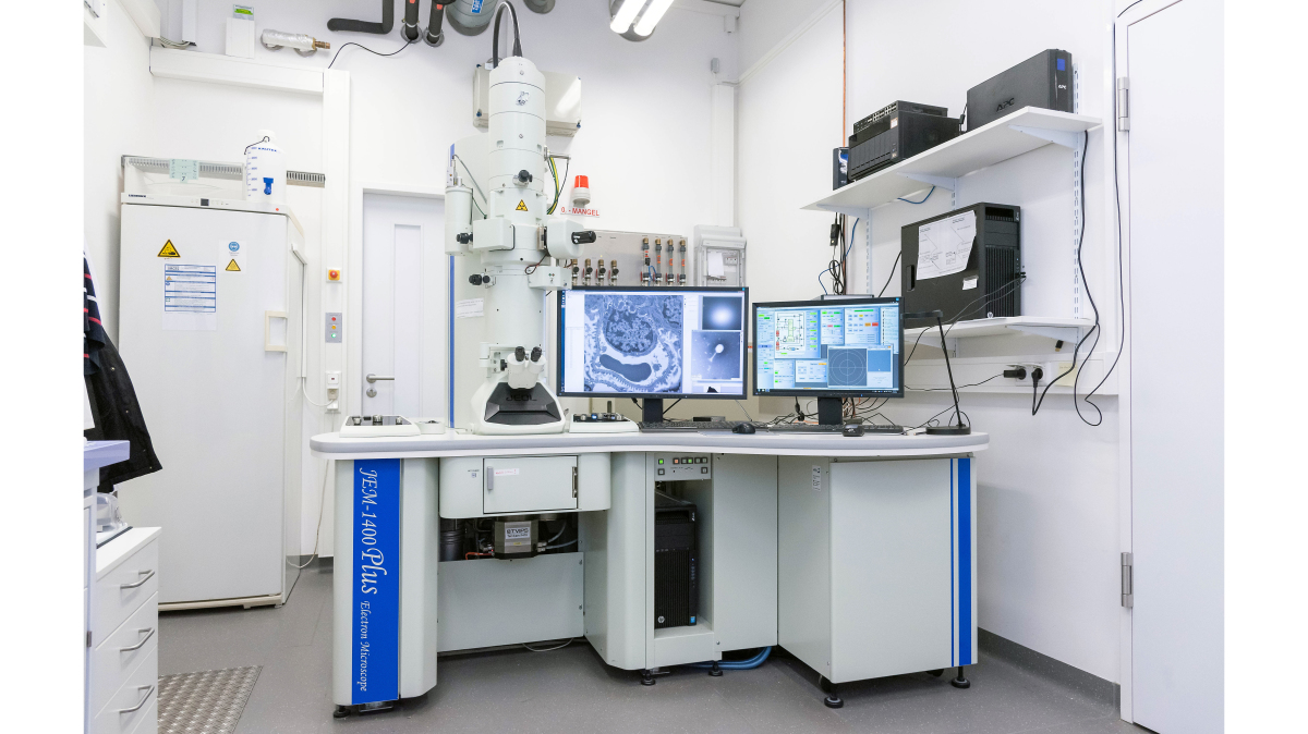
Room 1.035 1st floor MFZ building
Our transmission electron microscope allows looking through sectioned ultrastructures like cells and their organelles at a very high resolution. For example if you want to see if certain cell organelles, e.g., mitochondria are affected by certain experimental treatments in number or morphology transmission electron microscopy is the appropriate choice since it can visualise small structures by far better than light microscopy due to its resolution which is about 1,000x higher (point resolution 0.38 nm). By means of our ±70° tilt-holder the TEM allows to take tomography series of tiny structures like synapses for later 3D-reconstruction.
Equipped with a high contrast objective-poleshoe and a LaB6 Filament it is possible to perform cryo-electron microscopy using our cryo-specimen holder. In this context we can investigate ultra-quick frozen particles, e.g. viruses, by means of our plunge freezer avoiding fixation and embedding artifacts of conventional electron microscopy.
The integrated TVIPS TemCam-F416 high-sensitive CMOS camera allows to take 16Bit digital micrographs at a resolution of 4,096 x 4,096 pixels using the EM-Menu 4.0 extended software.
For long-term projects it is possible to work alone on the TEM after an appropriate instruction.