New discovered
Blood Vessel System in Bones
- von Milena Hänisch
- 21.01.2019
A previously unknown network of fine capillaries directly connecting the bone marrow with the circulation of the periosteum has been discovered by a team of scientists led by Prof. Matthias Gunzer and Dr. Anja Hasenberg from the Institute for Experimental Immunology and Imaging at the University Hospital of the University Duisburg-Essen (UDE) in Germany.
The group was further supported by research institutes in Erlangen, Jena, Berlin, Dresden and Berne (Switzerland). Their results have now been published in the prestigious international journal “Nature Metabolism”.
Bones are very hard organs. Still they do possess a tight meshwork of blood vessels in their inner cavity, where the bone marrow is located, as well as on the bone surface, that is covered by the highly vascularized periosteum. This is why e.g. bone fractures can bleed extensively. The same blood vessel system is also essential to transport blood- and immune cells from their place of origin, the bone marrow, to the outside.
„Like any other organ also bones need a closed circulatory loop (CCL) to function properly. This delivers fresh blood via arteries into the bone and transports used blood out via veins. How exactly the CCL of long bones functions was not totally clear until now.” says Dr. Anika Grüneboom from the University Hospital in Erlangen, first author of the study. She performed her PhD thesis in the group of Prof. Gunzer, where she completed most of the work, that has now been published.
Sometimes more than 1,000 capillaries per bone
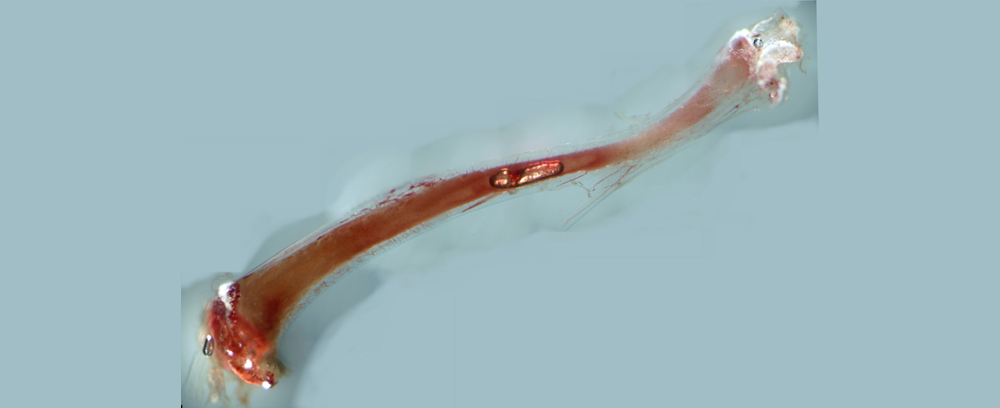
In the long bones of mice, the team observed and characterized a new type of capillary that perpendicularly to the long axis crosses the entire hard bone, which is termed corticalis. Hence the new blood vessels were named “trans cortical vessels” (TCV) and the team found hundreds to more than 1,000 of those in e.g. a mouse tibia. The vessels could either be of arterial or venous origin. Surprisingly, the scientists could demonstrate, that the overwhelming majority of arterial and venous blood in long bones flows through the TCV system.
Prof. Gunzer: „Previous concepts described just a handful of arterial inlets and two venous exits on long bones of mice. This is obviously too oversimplified and does not correctly reflect the true natural situation. It is really unexpected being able to find a new and central anatomical structure that has not been described in any textbook in the 21st century.”
This discovery was enabled by a unique combination of cutting edge imaging approaches that have been established and honed over many years. “Several of those methods have been used for the first time to study blood flow in bones.” says Prof. Gunzer. „This applies e.g. to the so-called Light sheet microscopy and 7 Tesla ultra high field magnetic resonance imaging.”
Research requiring full physical involvement
Using these techniques, it could be shown that TCVs also exist in human bones, at least in some parts of these much thicker structures. Thereby the study required full – also physical - involvement from all participants. Prof. Gunzer himself was lying in the 7 Tesla machine of the Erwin L. Hahn Institute at the UDE for ~6h until all necessary high-quality scans of his lower leg were in the box.
Future work shall now investigate which role is played by TCVs in normal bone physiology as well as under disease conditions such as osteoporosis, rheumatoid arthritis or tumors, that metastasize into bones.
Additional Information:
https://www.nature.com/articles/s42255-018-0016-5
Prof. Matthias Gunzer, Institute for Experimental Immunology and Imaging, Tel. 0201/18 3-6640, matthias.gunzer@uni-due.de