Decomposition of Damaged Lysosomes
New Signaling Pathway Decoded
- von Birte Vierjahn
- 08.04.2024
Lysosomes are the recycling centers in our cells: The spherical organelles break down both endogenous and foreign substances in their insides – for further utilization. As their interior has an acidic pH value, damage to the membrane surrounding the lysosomes is dangerous for cells. Scientists at the University of Duisburg-Essen have identified a new signaling pathway that in this case leads to the decomposition of the lysosome. Their findings, published in Molecular Cell, could contribute to the development of new approaches for the treatment of neurodegenerative diseases.
Lysosomes are surrounded by a lipid bilayer that separates the acidic environment and the digestive enzymes of the organelle from the cytoplasm. Damage to this layer – lysosomal membrane permeabilization, or LMP for short – can trigger inflammation and even lead to cell death. In humans, LMP is particularly deleterious in nerve cells during aging, inflammation or trauma because nerve cells cannot easily regenerate; however, intentionally inducing LMP in cancer cells can also be a therapeutic option.
In the event of membrane damage to a lysosome, a cell has two options: attempt a repair or disintegrate the organelle under safe conditions. How this decision is made is not yet fully understood. Scientists from Prof Dr Hemmo Meyer's research group at the University of Duisburg-Essen (UDE), together with colleagues from Munich and Milan, have investigated the reaction of cells to damage in lysosomal membranes. They were able to identify a previously unknown signaling pathway in human cells that is driven by the protein SPG20. It recognizes damage to the outer side of the lysosomal membrane, binds to the leaky organelle and triggers its breakdown before the membrane ruptures and the survival of the cell is endangered.
"Our findings could help to better understand the cellular response to lysosome damage and to develop new approaches for the treatment of neurodegenerative diseases associated with lysosome damage," says Pinki Gahlot, PhD student in molecular biology and first author of the publication. "Conversely, certain cancer cells seem to be particularly vulnerable to LMP – sp intentionally blocking the discovered factors could be a strategy in cancer therapy."
In further studies, the team wants to explore how the novel pathway is controlled by the cell to ensure cellular survival. For this purpose, they will use the new high-tech equipment in the university’s microscopy facility.
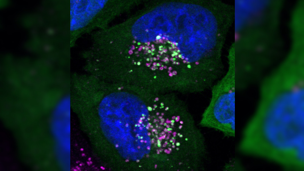
Image: The green dots are SPG20 which co-localizes with lysosomes represented in magenta. The nucleus of the cell is shown in blue.
Original Publication: https://doi.org/10.1016/j.molcel.2024.02.029
Further Information:
Prof. Dr. Hemmo Meyer, Molecular Biology I, +49 201/18 3-4217, hemmo.meyer@uni-due.de
Editor: Birte Vierjahn, +49 203/37 9-2427, birte.vierjahn@uni-due.de